Body Fluids, Blood, Lymphatic System & Inflammation
Blood is the liquid lifeline of the human body, transporting oxygen, nutrients, antibodies, and hormones to trillions of living cells. In this unit, we explain body fluid compartments, blood composition, hemopoiesis, the coagulation cascade, ABO/Rh blood grouping, the lymphatic defense system, the cellular mechanisms of acute inflammation, and common haematological diseases (anaemias and haemophilia).
In this topic, you will learn:
1. Body Fluid Compartments and Composition of Blood
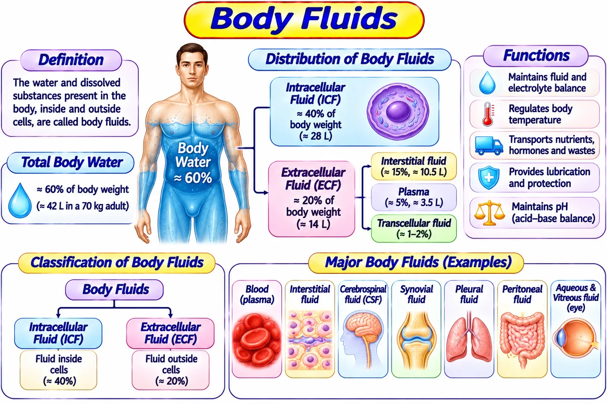
Visual Diagram: Total Body Water (TBW) Distribution in a 70 kg Adult (~42 Litres)
Fluid located inside all cells. Major cation is Potassium (K⁺); major anions are Phosphate and Proteins.
Fluid outside cells. Major cation is Sodium (Na⁺); major anion is Chloride (Cl⁻). Divided into:
Composition and Functions of Blood:
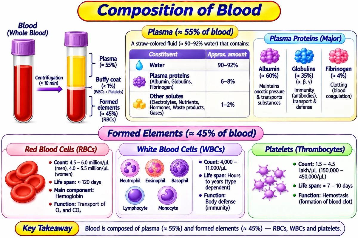
Blood is a specialized fluid connective tissue (total volume ~5 Litres, pH 7.35–7.45) consisting of:
Pale straw-colored liquid containing 91.5% water and 8.5% solutes:
- Serum Albumin (54%): Generates colloid osmotic pressure (25 mmHg) to keep fluid inside capillaries.
- Globulins (38%): Alpha/Beta transport lipids; Gamma globulins are protective antibodies (Immunoglobulins).
- Fibrinogen (7%): Soluble plasma precursor converted to insoluble fibrin clot during bleeding.
- Erythrocytes (RBCs):
• It is also known as: Red Blood Cells (RBCs).
• It is used for: Binding and transporting oxygen (O₂) to living tissues and carrying back carbon dioxide (CO₂) via Hemoglobin (lifespan = 120 days). - Leukocytes (WBCs):
• It is also known as: White Blood Cells (WBCs) / Cellular defense soldiers.
• It is used for: Protecting the body against viral, bacterial, and fungal infections, and foreign toxins:
• Granulocytes: Neutrophils (60–70% bacterial phagocytes), Eosinophils (allergies & parasites), Basophils (histamine release).
• Agranulocytes: Lymphocytes (T-cells & B-cells), Monocytes (macrophages). - Thrombocytes (Platelets):
• It is also known as: Blood platelets.
• It is used for: Initiating primary hemostasis by forming platelet plugs and activating the clotting cascade to stop bleeding.
2. Hemopoiesis and Formation of Hemoglobin
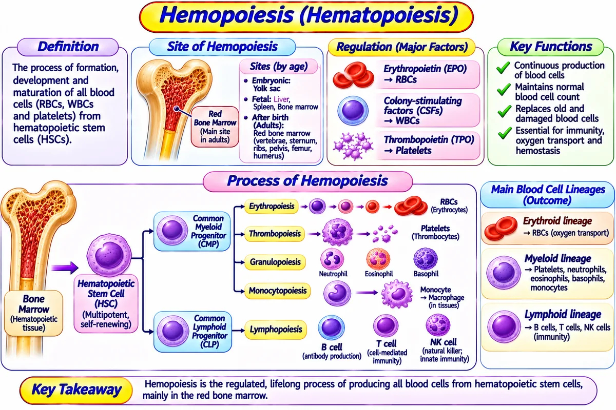
Simple Definition:
Hemopoiesis (Hematopoiesis) is defined as the physiological process of blood cell generation taking place primarily inside the red bone marrow of flat and long bones under the control of the hormone Erythropoietin (EPO) produced by the kidneys in response to hypoxia.
Stages of Erythropoiesis (RBC Maturation):
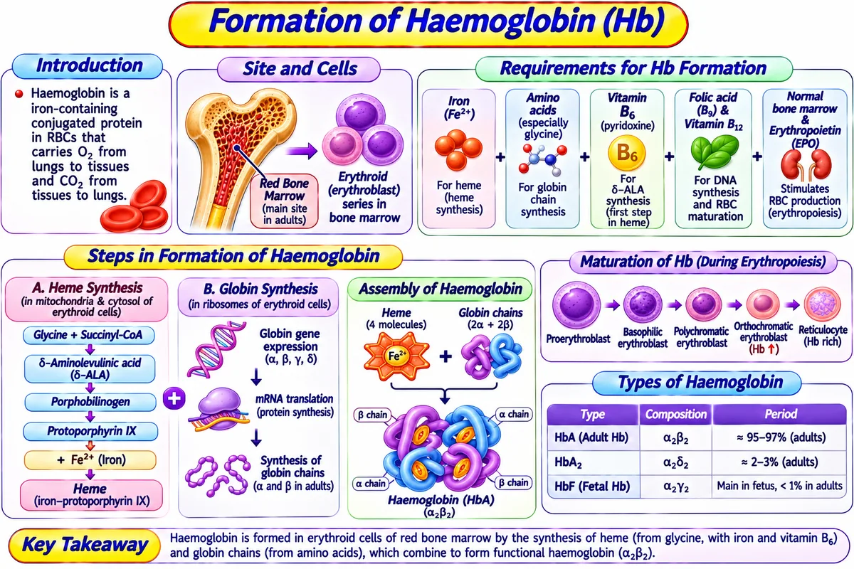
Structure and Synthesis of Hemoglobin (Hb):
Hemoglobin is a conjugated chromoprotein (normal adult concentration = 13–17 g/dL in males, 12–15 g/dL in females) consisting of:
- Globin Protein (96%): 4 polypeptide chains (Adult HbA has 2 Alpha & 2 Beta chains).
- Heme (4%): 4 iron-protoporphyrin IX complexes. Each heme contains one ferrous iron (Fe²⁺) atom at its center capable of loosely and reversibly binding one molecule of oxygen (O₂). Thus, 1 molecule of Hb carries exactly 4 molecules of O₂.
3. Mechanisms of Blood Coagulation (The Clotting Cascade)
Simple Definition:
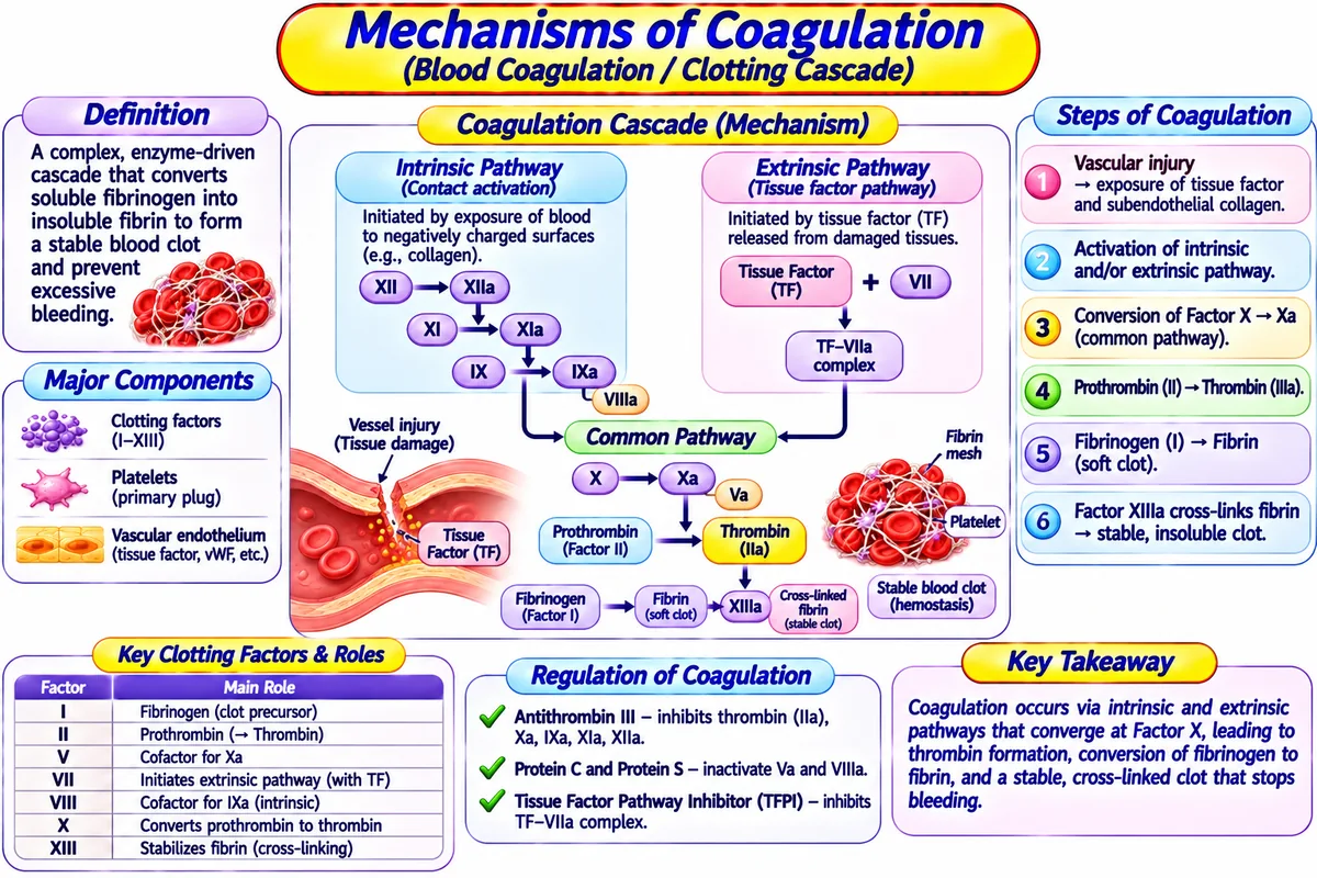
Blood Coagulation (Clotting) is defined as a complex enzymatic cascade of 13 plasma clotting factors whereby liquid blood solidifies into an insoluble fibrin meshwork to prevent fatal hemorrhage after vascular injury.
Visual Diagram: The Dual Pathway of Blood Coagulation
Triggered by trauma inside the blood vessel or contact with foreign glass/collagen → Factor XII activated → XIa → IXa + VIIIa complex activates Factor X.
Triggered by tissue damage outside vessel releasing Tissue Factor (Factor III / Thromboplastin) → complex with VIIa activates Factor X.
Factor Xa + Factor Va + Ca²⁺ (Factor IV) + Platelet Phospholipids → Prothrombinase Complex
Prothrombin (Factor II) → Thrombin (Factor IIa)
Fibrinogen (Factor I) → Insoluble Fibrin Monomers → Crosslinked by Factor XIIIa into Permanent Clot
4. Blood Grouping (ABO & Rh Factors) and Transfusion Principles
ABO Blood Grouping System & Agglutination
Simple Definition:
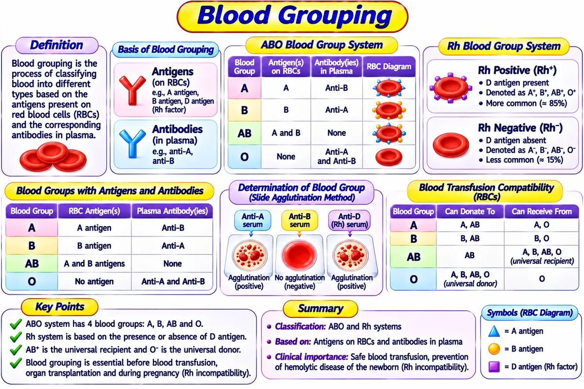
Blood Grouping is defined as the scientific system of classifying human blood based on the presence or absence of specific inherited antigens (agglutinogens) on the surface of red blood cells.
- • It consists of two major systems: 1) The ABO Blood Group System (Groups A, B, AB, O), and 2) The Rh Blood Group System (Rh-positive and Rh-negative).
- • It is used for: Safe blood transfusions, organ transplantation, and preventing hemolytic reactions.
Karl Landsteiner discovered that the membrane of human RBCs contains genetically determined surface glycoproteins called Agglutinogens (Antigens), while blood plasma contains natural matching Agglutinins (Antibodies):
| Blood Group | Antigen on RBC Surface | Antibody in Plasma | Can Receive Blood From | Clinical Transfusion Status |
|---|---|---|---|---|
| Group A | Antigen A | Anti-B antibody | A, O | Safe donor to A, AB |
| Group B | Antigen B | Anti-A antibody | B, O | Safe donor to B, AB |
| Group AB | Both A & B Antigens | NONE (No antibodies) | A, B, AB, O | Universal Recipient (AB+) |
| Group O | NONE (No antigens) | Both Anti-A and Anti-B | O only | Universal Donor (O-) |
Rh Factor, Transfusion Incompatibility & Erythroblastosis Fetalis
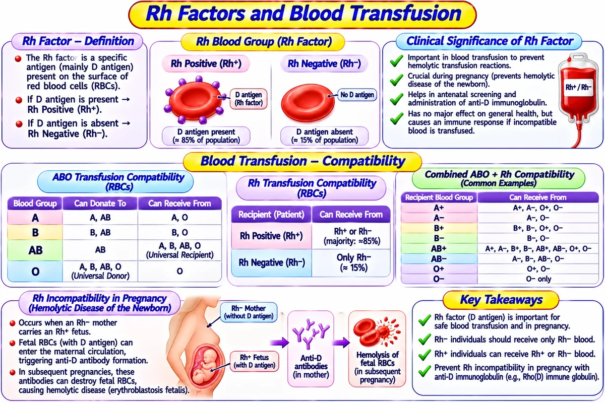
If an Rh-negative mother carries an Rh-positive fetus, fetal Rh+ RBCs enter maternal circulation during first delivery. The mother generates maternal Anti-Rh (anti-D) antibodies. In subsequent pregnancies with an Rh+ fetus, maternal IgG antibodies cross the placenta, destroying fetal RBCs causing fatal jaundice, hydrops fetalis, and kernicterus. Prevented by giving mother Anti-D Immunoglobulin (RhoGAM) within 72 hours of delivery.
5. Lymphatic Organs, Tissues, Lymph Formation and Circulation
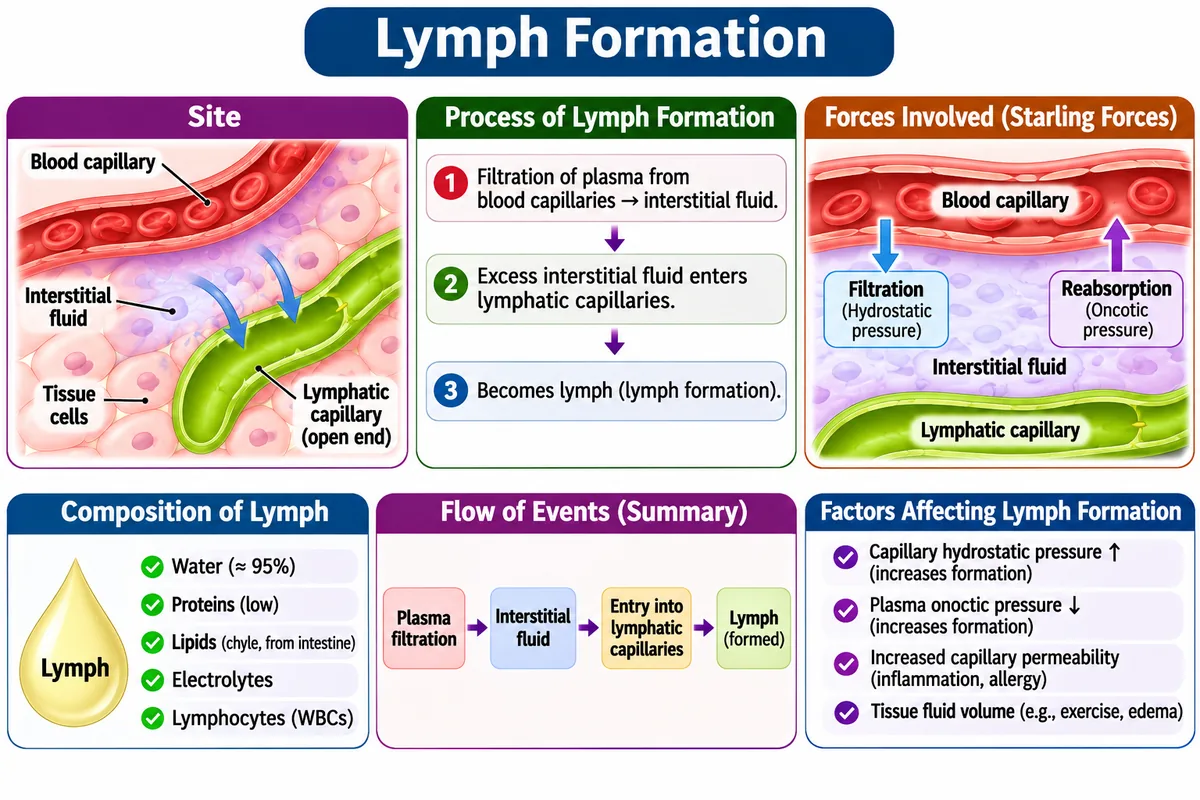
Lymph Formation and Interstitial Fluid Dynamics
Simple Definition:
The Lymphatic System is defined as a network of lymphatic vessels, lymph nodes, and lymphoid organs that drains excess interstitial fluid (~3 Litres/day) back into the venous bloodstream and houses immune cells to filter pathogens.
Primary & Secondary Lymphoid Organs (Thymus, Spleen, Lymph Nodes)
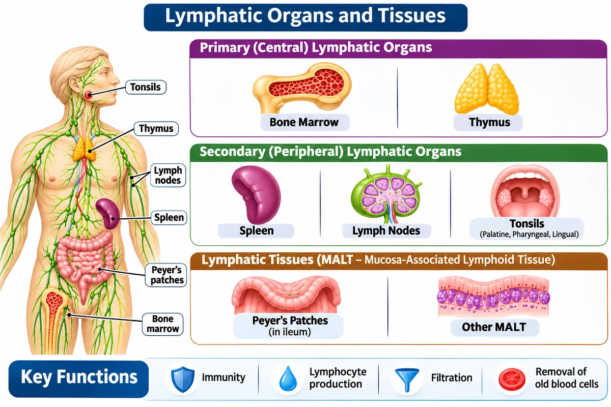
Filters blood, removes old fragile RBCs (graveyard of RBCs), stores blood platelets, and houses splenic B/T-lymphocytes.
Bean-shaped filters along vessels; contain macrophages and lymphocytes that destroy trapped bacteria and metastatic cancer cells.
Located in anterior mediastinum; primary site where immature T-lymphocytes differentiate and mature for cell-mediated immunity.
Palatine tonsils and Mucosa-Associated Lymphoid Tissue (Peyer's patches in ileum) that trap swallowed or inhaled microbes.
6. Basic Mechanism and Chemical Mediators of Inflammation
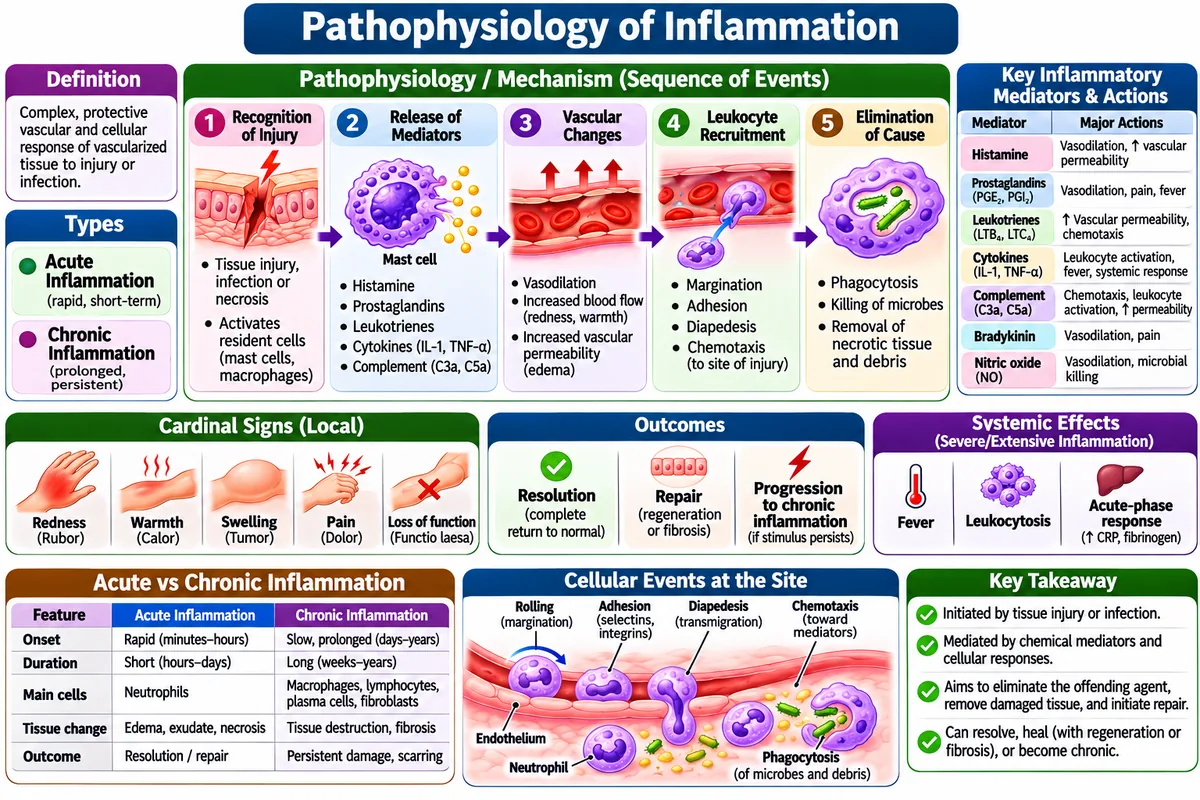
Simple Definition:
Inflammation is defined as a protective vascular connective tissue response elicited by the host body to destroy, dilute, or wall off invading pathogens, physical toxins, or necrotic tissue.
The 5 Cardinal Signs of Acute Inflammation (Celsus & Virchow):
Key Chemical Mediators of Inflammation:
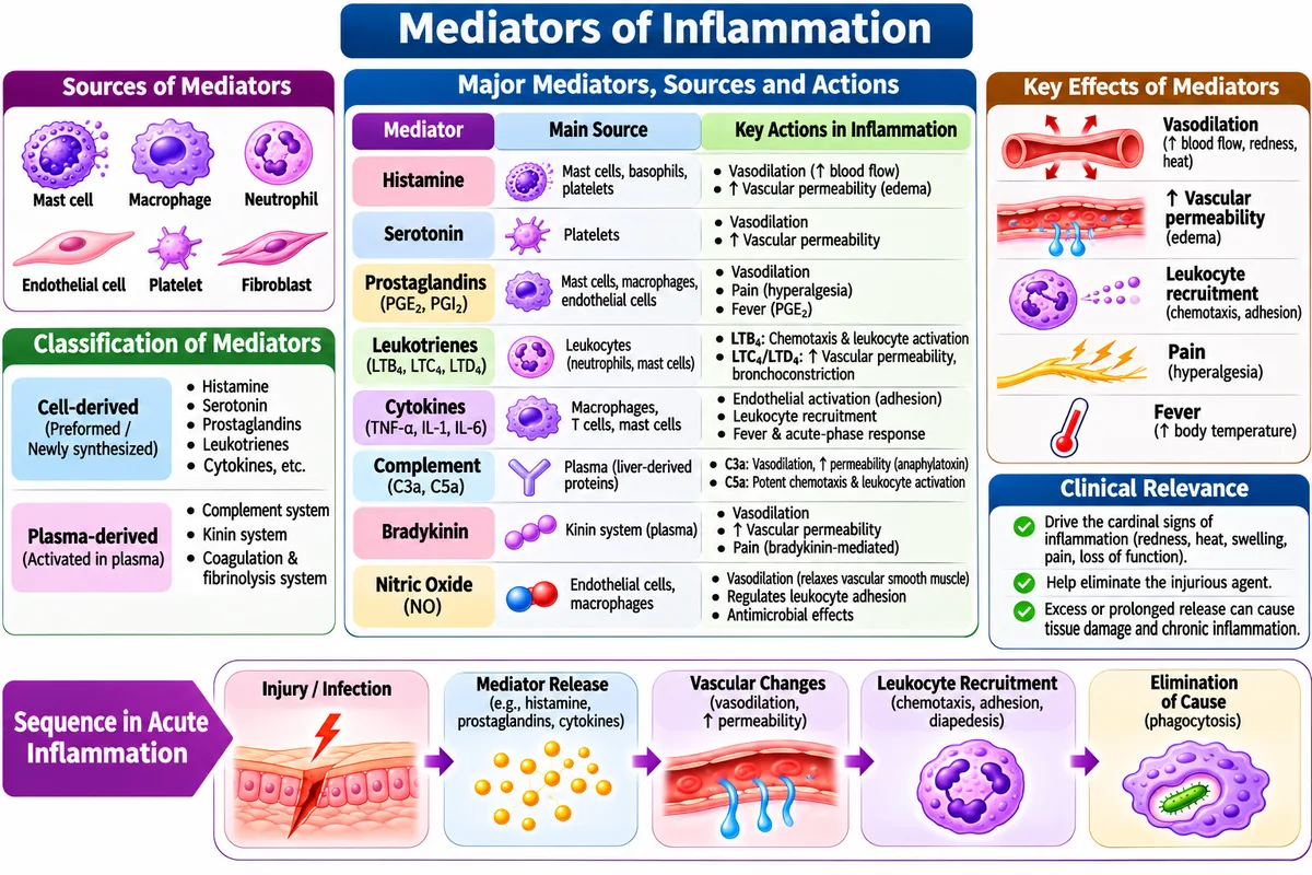
| Mediator | Origin / Source | Primary Inflammatory Action |
|---|---|---|
| Histamine | Mast cells and Basophils | Immediate arteriolar vasodilation and increased venular permeability. |
| Prostaglandins (PGE₂) | Cyclooxygenase (COX) enzyme from cell membranes | Vasodilation, potentiation of pain, and hypothalamic fever induction (inhibited by Paracetamol & NSAIDs). |
| Leukotrienes (LTB₄, LTC₄) | Lipoxygenase (LOX) pathway | LTB₄ is a powerful neutrophil chemoattractant; LTC₄/LTD₄ cause severe bronchoconstriction in asthma. |
| Bradykinin | Kallikrein-kinin plasma system | Intense pain receptor stimulation and smooth muscle contraction. |
7. Pathophysiology of Haematological Diseases: Anaemias and Haemophilia
1. Iron Deficiency Anaemia
Pathophysiology: Iron deficiency is the most common nutritional anaemia worldwide. Lack of dietary iron or chronic occult blood loss impairs heme synthesis, resulting in small, pale red blood cells (Microcytic, Hypochromic anaemia). Diagnostic findings include reduced serum iron, depleted serum ferritin, and elevated total iron-binding capacity (TIBC).
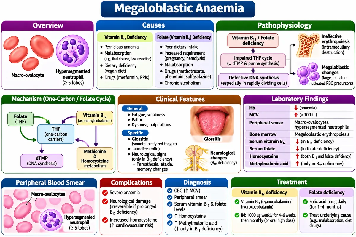
2. Megaloblastic Anaemia (Vitamin B₁₂ & Folate Deficiency)
Pathophysiology: Deficiency of either Vitamin B₁₂ (Cobalamin) or Folic Acid (Vitamin B₉) impairs thymidine nucleotide synthesis required for cellular DNA replication. Nuclear maturation lags behind cytoplasmic RNA and protein synthesis, leading to giant, fragile, abnormally shaped red blood cells (Macrocytic anaemia, with hypersegmented neutrophils). Pernicious anaemia is an autoimmune condition characterized by autoantibodies against gastric parietal cells or Intrinsic Factor, eliminating B₁₂ absorption in the terminal ileum.
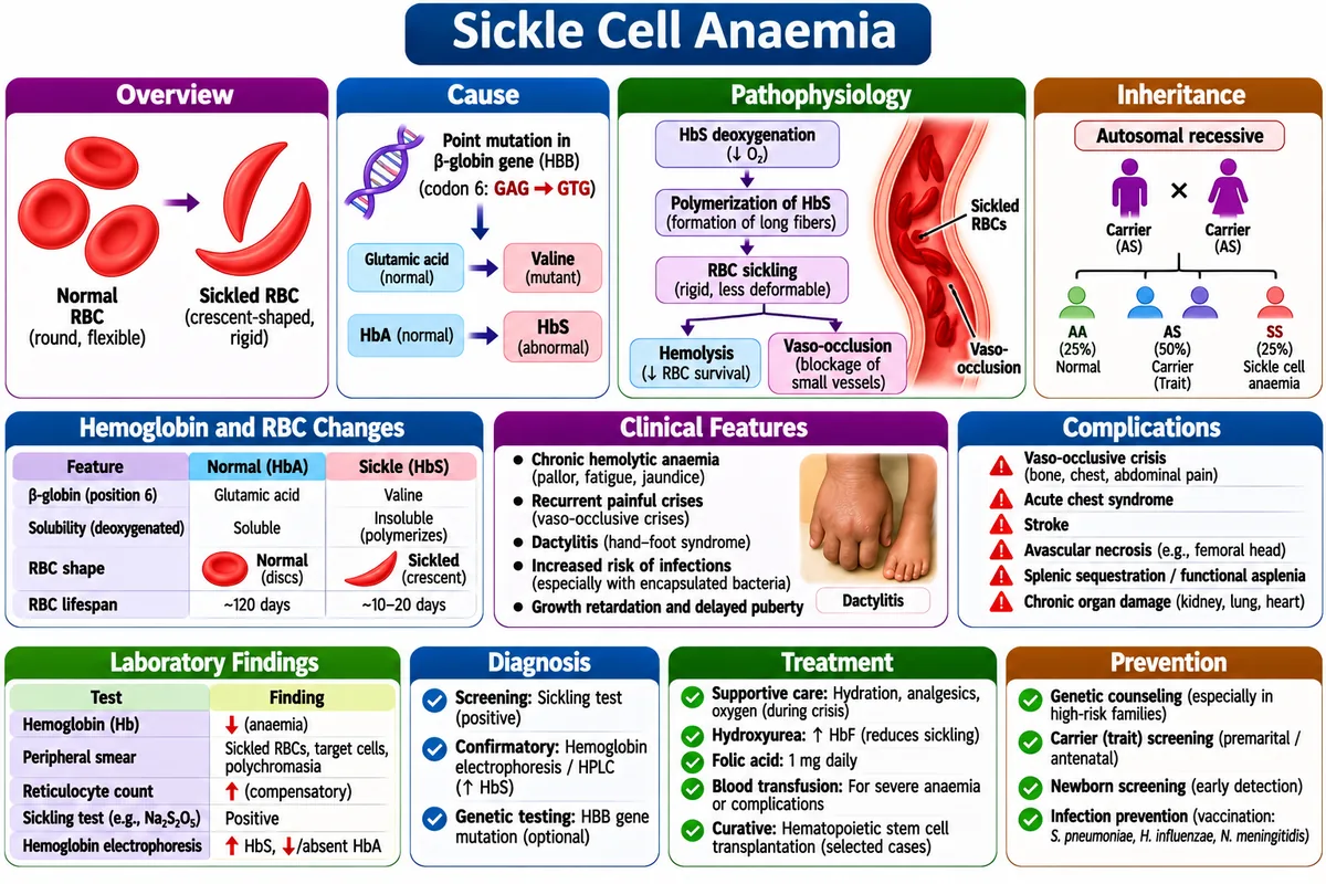
3. Sickle Cell Anaemia
Sickle Cell Anaemia is defined as an inherited autosomal recessive hemoglobinopathy caused by a point mutation in the beta-globin chain (HbS).
• It is also known as: Sickle cell disease (HbSS).
• It is used for: Explaining painful vaso-occlusive crises, hemolytic jaundice, and microvascular blockages caused by rigid sickle-shaped RBCs.
• In this mechanism, we: See valine replace glutamic acid at codon 6 of the beta-globin chain, causing hemoglobin polymerisation under low oxygen tension.
Pathophysiology: Sickle cell anaemia is an autosomal recessive genetic haemoglobinopathy caused by a single point mutation (missense mutation) in the β-globin gene: Valine substitutes for Glutamic acid at the 6th position of the β-polypeptide chain, creating abnormal Hemoglobin S (HbS). Under deoxygenated conditions (hypoxia, acidosis, dehydration), HbS molecules polymerize into long, stiff, insoluble fibers, distorting pliable round erythrocytes into rigid, fragile, crescent or sickle shapes.
4. Thalassemia (α and β Thalassemia)
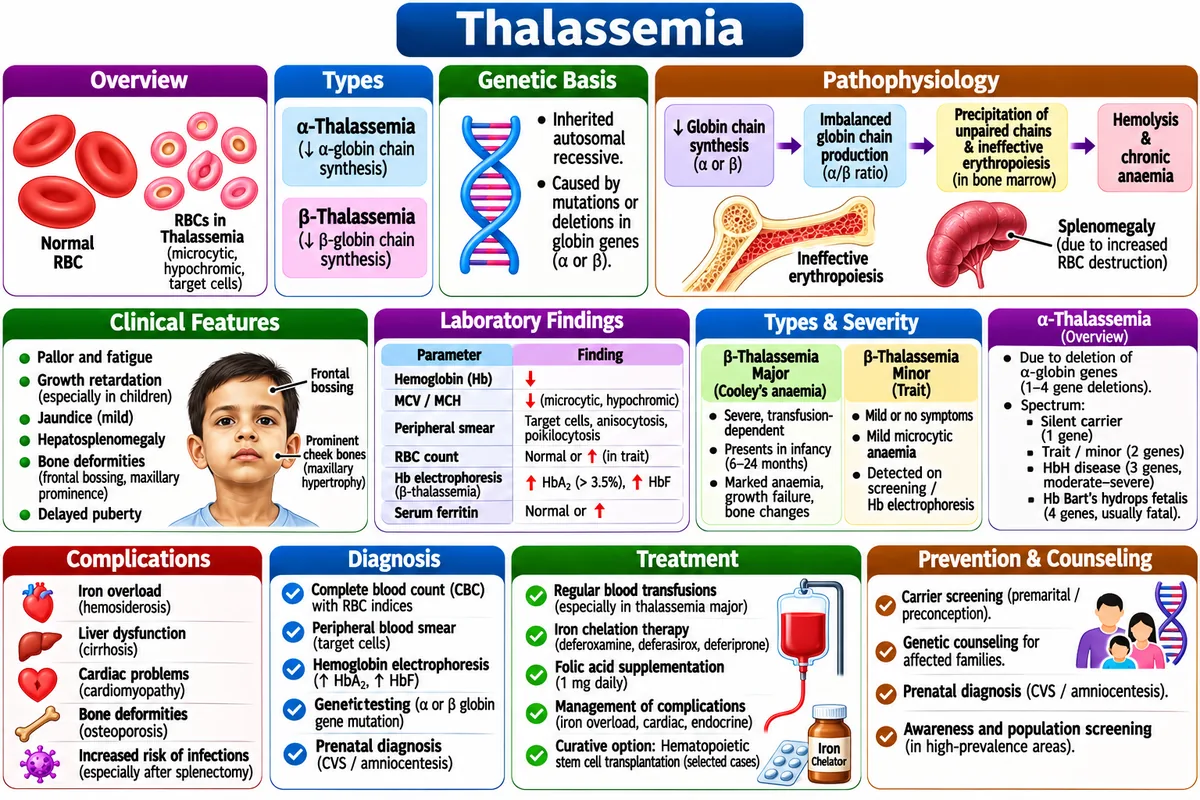
Thalassemia is defined as a hereditary microcytic hemolytic anemia characterized by absent or reduced synthesis of alpha or beta globin chains.
• It is also known as: Mediterranean anemia or Cooley's anemia (Beta Thalassemia Major).
• It is used for: Explaining severe chronic anemia requiring life-long blood transfusions and iron chelation therapy.
• For example: Unpaired excess globin chains precipitate inside erythroblasts, destroying them prematurely in bone marrow.
Pathophysiology: Thalassemia is a group of hereditary microcytic haemolytic anaemias caused by mutations or gene deletions that reduce or completely halt the synthesis of either α- or β-globin polypeptide chains of adult hemoglobin (HbA, α₂β₂). The resulting imbalance produces precipitating excess unmatched globin chains that damage RBC membranes, resulting in ineffective erythropoiesis inside bone marrow and premature peripheral hemolysis in the spleen.
5. Haemophilia (Haemophilia A & B)
Pathophysiology: Haemophilia is an X-linked recessive bleeding disorder predominantly affecting males, characterized by deficient intrinsic coagulation pathway activity:
- Haemophilia A (Classical Haemophilia, ~80–85% of cases): Congenital deficiency of Factor VIII.
- Haemophilia B (Christmas Disease, ~15–20% of cases): Congenital deficiency of Factor IX.