Cardiovascular System and Heart Disorders
The human heart beats approximately 100,000 times every single day, pumping over 7,000 litres of blood through 100,000 kilometers of blood vessels. In this vital unit, we cover the anatomy of heart chambers and valves, blood circulation routes, blood vessel histology, the cardiac conduction pacemaker system, the cardiac cycle and ECG waveforms, blood pressure regulation (including the RAAS pathway), and major cardiovascular diseases (hypertension, angina, myocardial infarction, atherosclerosis, and heart failure).
In this topic, you will learn:
1. Gross Anatomy of the Heart and Blood Circulation Routes
Simple Definition:
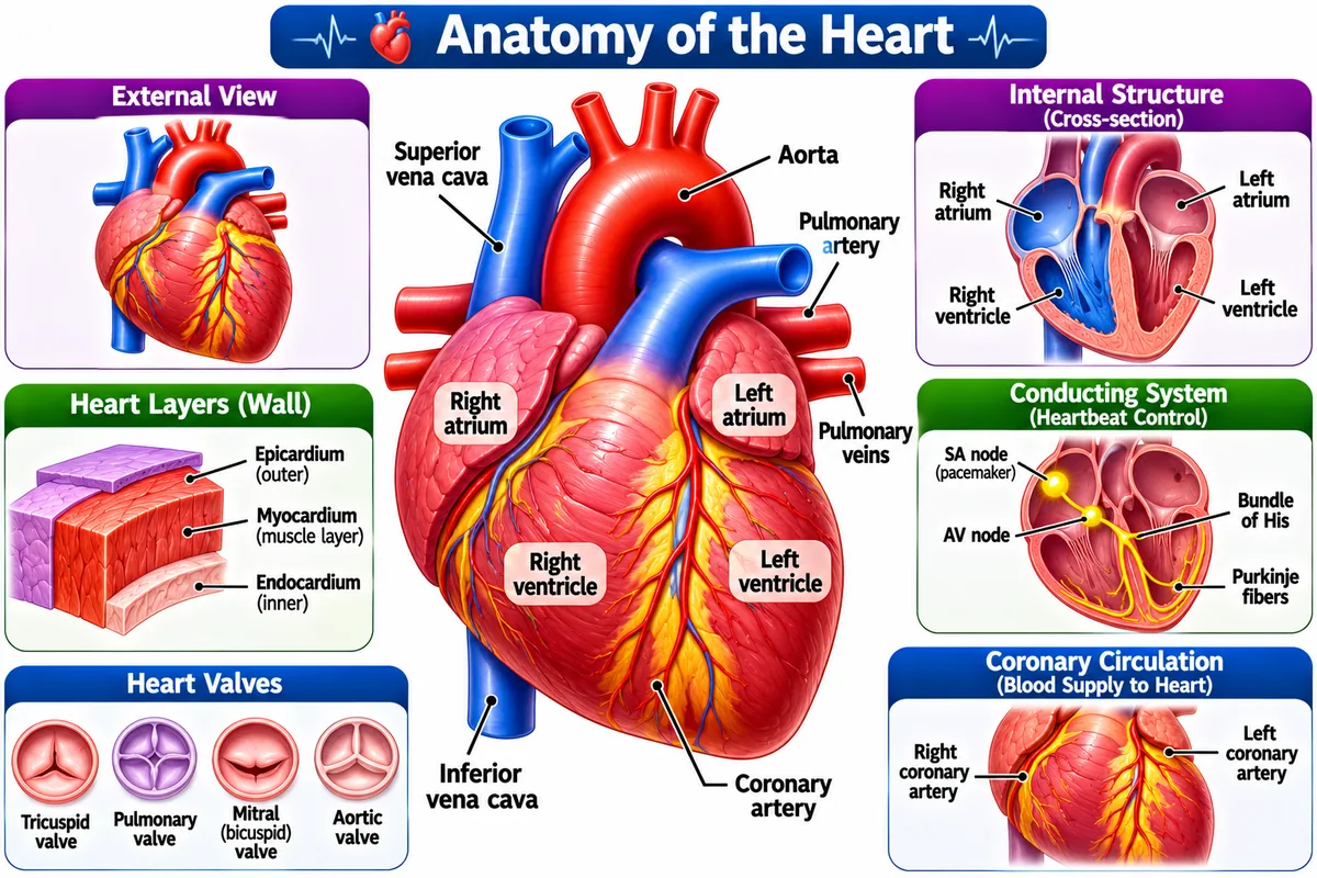
The Heart is defined as a hollow, muscular, four-chambered double pump roughly the size of a closed fist (weighing ~250–350 g), located in the thoracic mediastinum between the two lungs, tilted towards the left side.
- Right Atrium (RA): Receives deoxygenated venous blood from Superior/Inferior Vena Cava and Coronary Sinus.
- Right Ventricle (RV): Pumps deoxygenated blood into Pulmonary Artery toward lungs (low pressure).
- Left Atrium (LA): Receives oxygen-rich blood from 4 Pulmonary Veins.
- Left Ventricle (LV): Thickest muscular wall (3x thicker than RV); pumps oxygenated blood into Aorta to entire systemic body under high pressure (120 mmHg).
- Wall Layers: Outermost Epicardium → thick contractile Myocardium (cardiac muscle) → smooth innermost Endocardium.
- Tricuspid Valve: 3 cusps; located between Right Atrium and Right Ventricle.
- Bicuspid (Mitral) Valve: 2 cusps; located between Left Atrium and Left Ventricle.
- Pulmonary Semilunar Valve: Located at exit of Right Ventricle into Pulmonary Trunk.
- Aortic Semilunar Valve: Located at exit of Left Ventricle into Ascending Aorta.
- Tendon cords (Chordae Tendineae) anchored to Papillary muscles prevent AV valves from inverting during ventricular contraction.
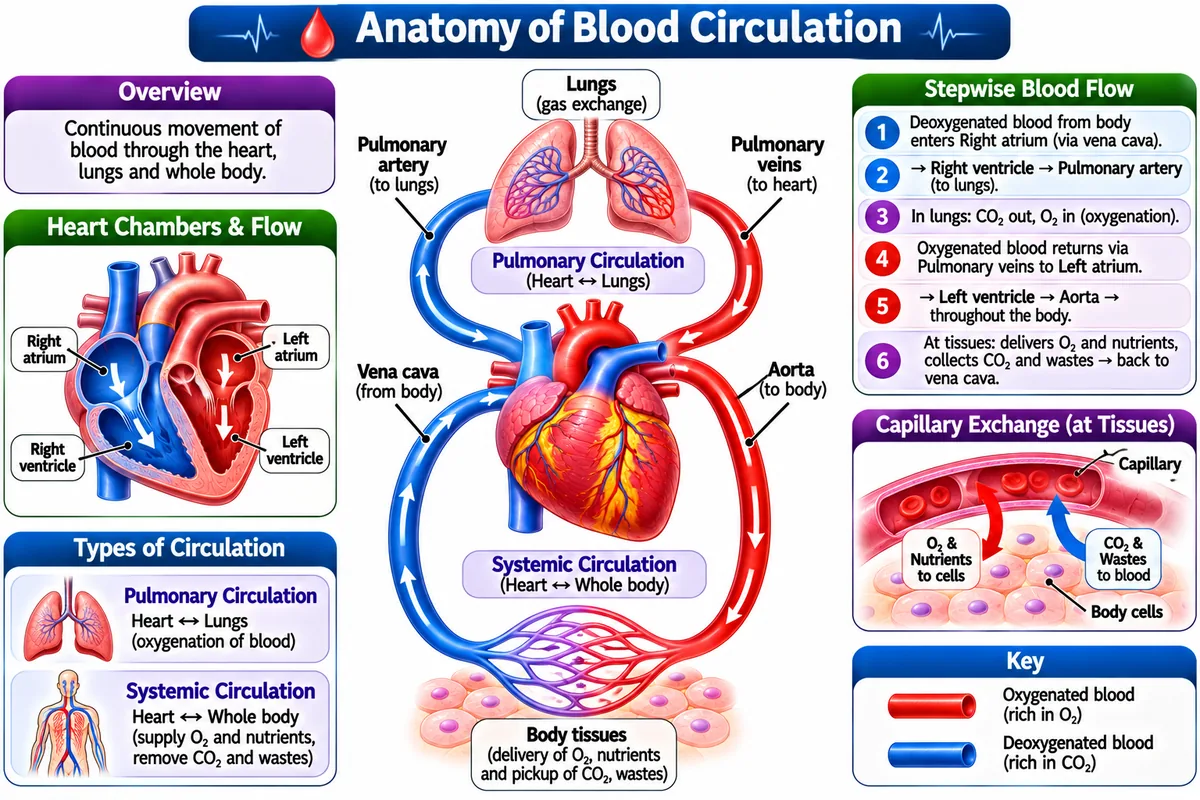
Visual Diagram: Complete Systemic & Pulmonary Blood Circulation Pathway
2. Blood Vessels: Structure and Functions of Arteries, Veins and Capillaries
Simple Definition:
Blood Vessels are defined as the closed network of muscular and elastic tubular conduits that carry blood from the heart to every tissue in the body and back to the heart.
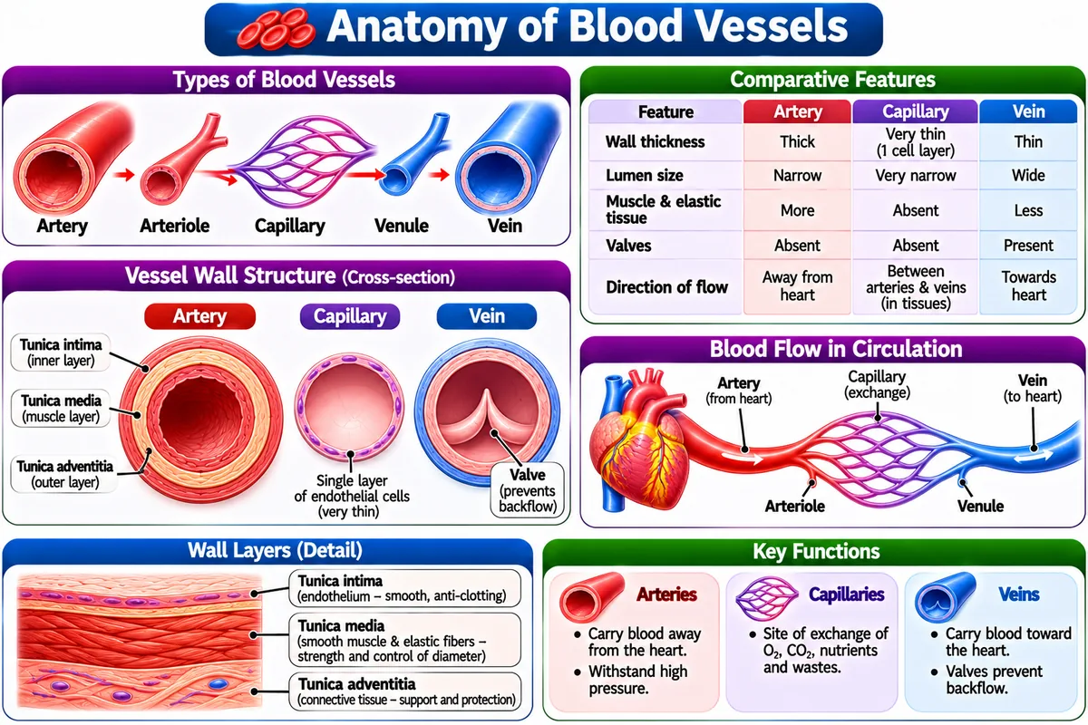
- • There are total (3) primary types of blood vessels: 1) Arteries (carry blood away from heart), 2) Veins (carry blood towards heart), and 3) Capillaries (microscopic vessels for gas and nutrient exchange).
- • It consists of three wall layers (Tunics): Tunica intima (inner endothelium), Tunica media (smooth muscle and elastic tissue), and Tunica externa (outer fibrous connective tissue).
- • It is responsible for: Maintaining continuous perfusion of blood, regulating blood pressure, and delivering O₂ and nutrients to cells.
| Vessel Type | Wall Histology (3 Tunics) | Pressure & Flow Direction | Key Distinguishing Features |
|---|---|---|---|
| Arteries | Thick muscular walls: very thick Tunica Media rich in smooth muscle and elastic lamina. | High pressure (80–120 mmHg); carry blood Away from heart. | Narrow lumen; no internal valves; expand and recoil with pulse waves (Aorta, Carotid). |
| Veins | Thin muscular walls; thin Tunica Media, thick Tunica Externa; wide lumen. | Low pressure (2–10 mmHg); return blood Toward the heart. | Contain one-way semilunar venous valves preventing backward pooling; act as blood capacitance reservoirs (store 64% of blood). |
| Capillaries | Microscopic single layer of Endothelial cells resting on thin basement membrane; no media or externa. | Slowest velocity (0.1 cm/s); connects arterioles to venules. | Sole site of metabolic exchange of oxygen, glucose, and wastes between blood and tissues. |
Microscopic Structure of an Artery
Arteries carry blood under high pulsating systolic pressure away from the heart. Their thick wall features an elastic internal and external lamina with abundant smooth muscle in the Tunica Media allowing dynamic vasoconstriction and vasodilation.
Architecture of Veins & One-Way Valves
Veins are thin-walled capacitance vessels that store up to 64% of total blood volume at low pressure. Because venous pressure is minimal, veins possess semi-lunar one-way pocket valves formed by foldings of the tunica intima to prevent backwards reflux of blood.
Capillary Bed and Microvascular Exchange
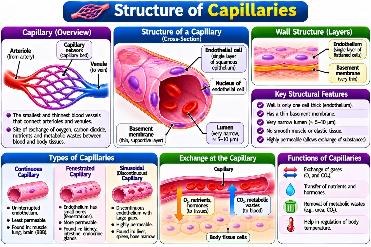
Capillaries consist solely of a single monolayer of simple squamous endothelial cells surrounded by a thin basement membrane (diameter ~5–10 μm), allowing rapid diffusion of gases, nutrients, and waste products between plasma and interstitial fluid.
3. Elements of the Conduction System of the Heart and Autonomic Regulation
Simple Definition:
The Cardiac Conduction System is defined as a network of specialized, non-contractile autorhythmic myocardial fibers that spontaneously generate and rapidly distribute rhythmic electrical action potentials to trigger coordinated heart contractions.
Visual Diagram: Electrical Impulse Conduction Pathway Through the Heart
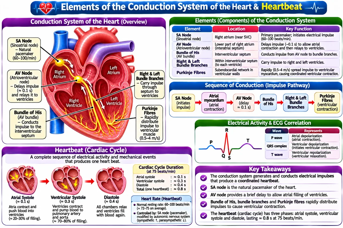
Located in wall of Right Atrium near SVC entrance. Fires spontaneously at 70–80 beats/min. Initiates each heartbeat wave spreading across atria.
Located in interatrial septum. Introduces an essential 0.1 second delay allowing atria to completely empty blood into ventricles before ventricles contract!
Only electrical bridge connecting atria to ventricles. Traverses down interventricular septum, dividing into Right and Left Bundle Branches.
Large, high-speed conduction fibers that curve upward from cardiac apex through ventricular walls, causing ventricles to contract upward toward aortic valve.
Autonomic Regulation of Heartbeat:
• Sympathetic Stimulation: Cardiac accelerator nerves release Noradrenaline acting on β₁ adrenergic receptors → increases SA node firing (tachycardia) and increases myocardial contractility.
• Parasympathetic Stimulation: The Vagus Nerve (Cranial Nerve X) releases Acetylcholine acting on M₂ muscarinic receptors → decreases SA node firing (bradycardia), slowing heart rate back to resting level.
4. Cardiac Output, Stroke Volume, Cardiac Cycle and Heart Sounds
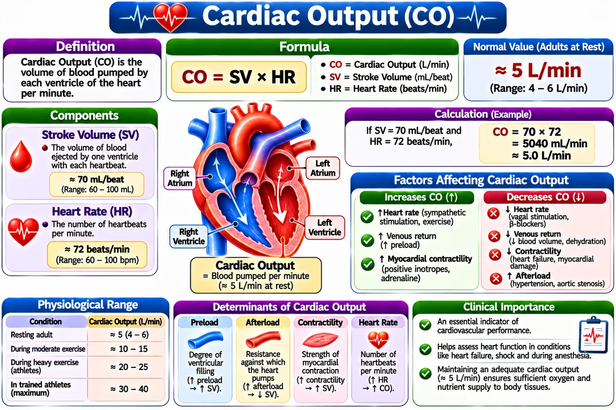
Cardiac Output (CO) Mathematical Formula:
Cardiac Output is defined as the total volume of blood ejected by each ventricle into the systemic or pulmonary circulation in one minute:
• Stroke Volume (SV): Volume ejected per beat ≈ 70 mL/beat.
• Resting Heart Rate (HR): ≈ 72 beats/minute.
Calculation: CO = 70 mL × 72 = 5,040 mL/min ≈ 5.0 Litres/minute (roughly entire blood volume circulating every minute!).
The 0.8 Second Cardiac Cycle & Heart Sounds:
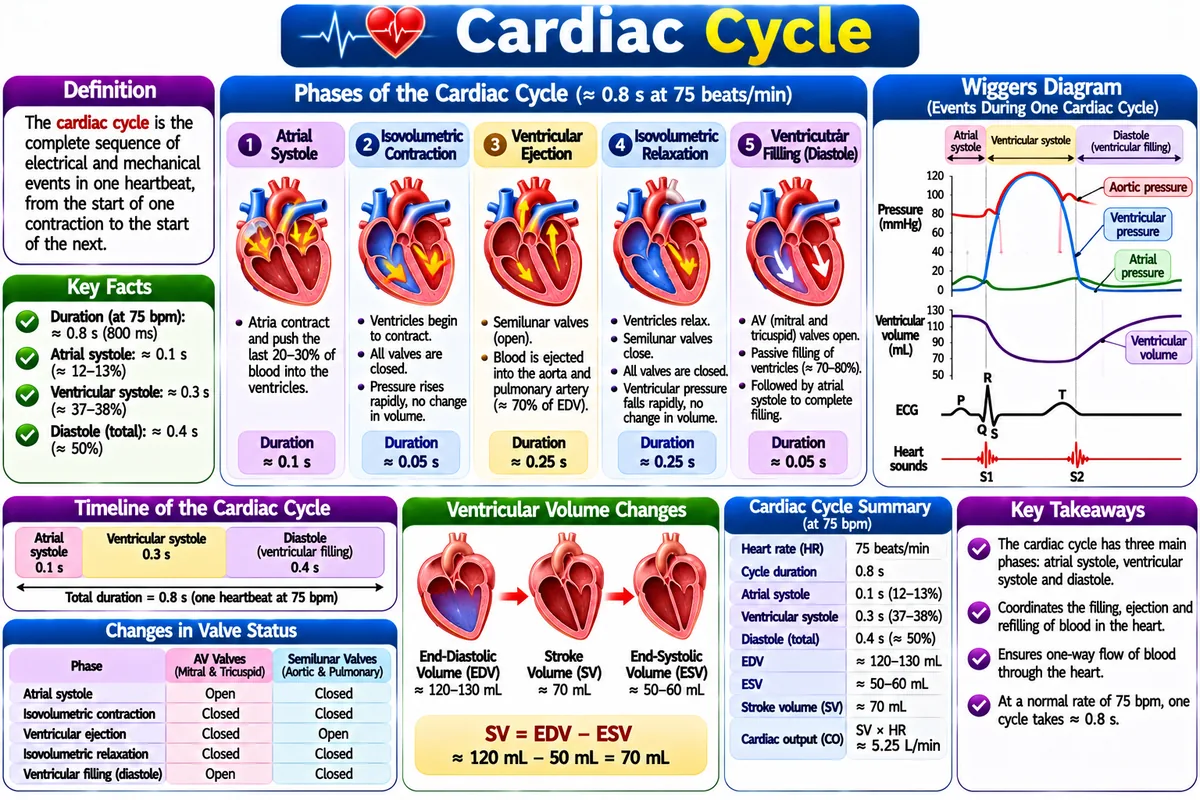
Produced by closure of the Atrioventricular (Tricuspid & Mitral) valves at the onset of ventricular systole. Long, soft, low-pitched sound.
Produced by closure of the Semilunar (Aortic & Pulmonary) valves at the onset of ventricular diastole. Sharp, short, high-pitched sound.
5. Regulation of Blood Pressure (RAAS Pathway) and the Electrocardiogram (ECG)
A. The Electrocardiogram (ECG / EKG): Deflections & Electrical Events
The Electrocardiogram (ECG) is defined as a graphical recording of the electrical potentials generated by muscle cells of the heart during each cardiac cycle, measured on the body surface using skin electrodes.
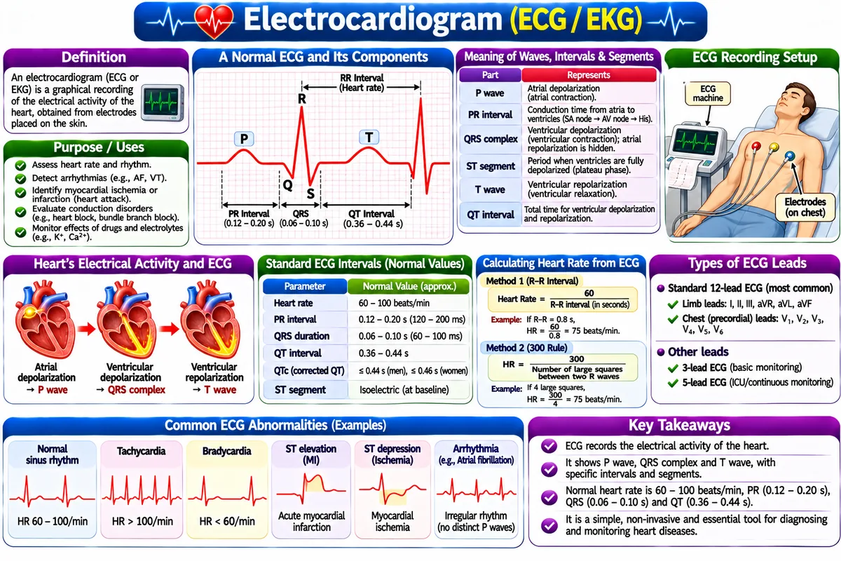
Spread of electrical impulse from SA node across atria, triggering atrial contraction.
Rapid spread of action potential down Bundle of His and Purkinje fibers; triggers ventricular contraction (atrial repolarization hidden inside).
Electrical recovery and relaxation of ventricles just before diastole.
B. Blood Pressure Regulation: Neural Reflexes & The RAAS Hormone Cascade
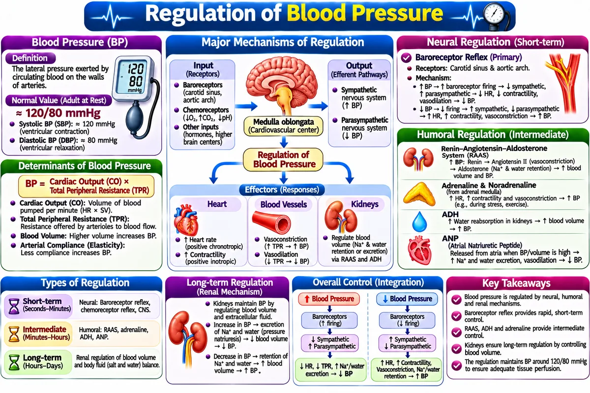
When blood pressure or renal blood flow drops, the kidneys activate the Renin-Angiotensin-Aldosterone System (RAAS):
1. Juxtaglomerular kidney cells secrete the enzyme Renin into the bloodstream.
2. Renin converts plasma Angiotensinogen (from liver) → Angiotensin I.
3. ACE (Angiotensin Converting Enzyme) in pulmonary capillaries converts Angiotensin I → Angiotensin II.
4. Angiotensin II Actions:
• Potent direct systemic Vasoconstriction (rapidly raises BP).
• Stimulates Adrenal Cortex to release Aldosterone (causes kidney tubules to retain Na⁺ and water, expanding blood volume).
Result: Total blood volume expands and vascular resistance increases → Blood Pressure is restored to normal! (Inhibited clinically by ACE inhibitors like Enalapril).
6. Pathophysiology of Cardiovascular Diseases: Hypertension, Angina, MI & CHF
1. Hypertension (High Blood Pressure)
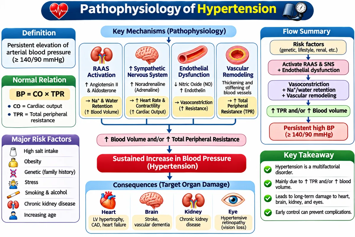
Hypertension is defined as persistent, sustained elevation of systemic arterial blood pressure above 140/90 mmHg.
• It is also known as: The silent killer.
• It is used for: Assessing cardiovascular risk and directing antihypertensive drug therapies.
• For example: A persistent reading of 150/95 mmHg requires lifestyle modification and ACE inhibitors.
Pathophysiology: Hypertension is defined clinically as persistent, sustained elevation of systemic arterial blood pressure: Systolic Blood Pressure ≥ 140 mmHg or Diastolic Blood Pressure ≥ 90 mmHg on repeated clinical measurements. Primary (essential) hypertension accounts for 90–95% of cases and arises from an interplay of genetic factors, sympathetic overactivity, endothelial dysfunction, and excess renal sodium reabsorption leading to increased total peripheral resistance (TPR). Secondary hypertension (5–10%) is caused by identifiable conditions such as renal artery stenosis, chronic kidney disease, pheochromocytoma, or Cushing's syndrome.
2. Atherosclerosis & Coronary Artery Disease (CAD)
Pathophysiology: Atherosclerosis is a chronic, progressive inflammatory disease affecting large and medium muscular elastic arteries. Endothelial injury triggered by hypertension, smoking, hyperlipidemia, and diabetes allows circulating Low-Density Lipoproteins (LDL) to infiltrate the tunica intima. Free radical oxidation of LDL recruits circulating monocytes, which differentiate into macrophages. Macrophages ingest oxidized LDL via scavenger receptors, transforming into lipid-laden foam cells.
3. Angina Pectoris (Ischemic Heart Disease)
Angina Pectoris is defined as sudden, suffocating substernal chest discomfort caused by transient myocardial ischemia without cellular death.
• It is also known as: Cardiac ischemic chest pain.
• It is used for: Warning patients of coronary artery narrowing before irreversible heart muscle death occurs.
• For example: Exertional chest tightness relieved within minutes by sublingual nitroglycerin tablets.
Pathophysiology: Angina pectoris is the clinical manifestation of transient myocardial ischemia resulting from a critical mismatch between myocardial oxygen demand (governed by heart rate, inotropy, and wall tension) and coronary blood oxygen supply. Oxygen starvation forces cardiomyocytes into anaerobic glycolysis, accumulating lactic acid and adenosine that irritate cardiac sympathetic sensory nerve endings (C7–T4), causing severe substernal chest discomfort radiating to the left shoulder, inner arm, neck, or jaw.
Predictable chest pain triggered by physical exertion or emotional stress; relieved within minutes by rest or sublingual Nitroglycerin.
Pain occurs at rest, with increasing frequency or severity; reflects acute plaque fissure with non-occlusive thrombus; pre-infarction medical emergency.
Episodic chest pain occurring at rest, caused by severe focal coronary artery vasospasm; treated with Calcium Channel Blockers.
4. Myocardial Infarction (MI / "Heart Attack")
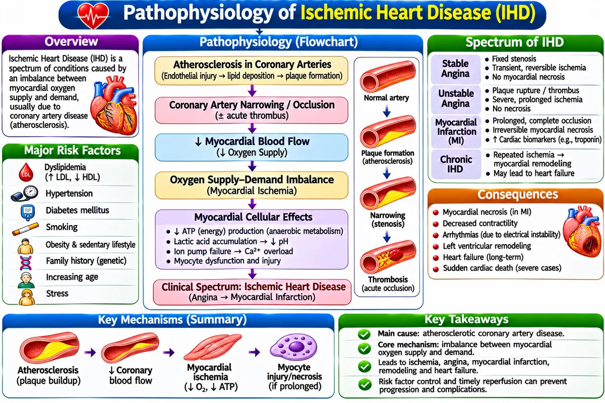
Myocardial Infarction (MI) is defined as irreversible ischemic necrosis of cardiac myocytes caused by prolonged acute coronary artery occlusion.
• It is also known as: Heart attack or acute coronary syndrome.
• It is used for: Identifying critical emergencies requiring immediate thrombolytic clot dissolution (e.g. Streptokinase) or angioplasty.
• For example: Crushing substernal chest pain radiating to left arm and jaw with ST-segment elevation on ECG.
Pathophysiology: Myocardial infarction occurs when an unstable atheromatous plaque ruptures, exposing pro-coagulant collagen and tissue factor to blood. Platelets rapidly adhere, aggregate, and generate an occlusive fibrin thrombus that completely blocks coronary arterial blood supply. Cardiomyocytes deprived of oxygen deplete ATP within minutes; irreversible ischemic necrosis (coagulative necrosis) begins within 20–30 minutes, starting from the vulnerable subendocardium and spreading transmurally toward the epicardium.
• Cardiac Troponins (cTnI and cTnT): Gold standard; rise within 3–4 hours, peak at 24 hours, and stay elevated for up to 10–14 days.
• CK-MB: Isoenzyme of creatine kinase; peaks at 24 hours and normalizes within 48–72 hours (useful for detecting re-infarction).
• ECG Findings: ST-segment elevation (STEMI: transmural infarction) or ST-depression / T-wave inversion (NSTEMI: subendocardial infarction) followed by pathological Q-waves.
5. Congestive Heart Failure (CHF)
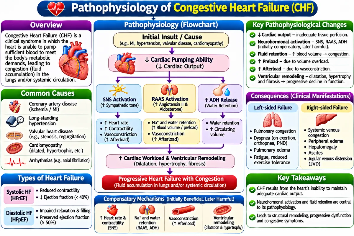
Congestive Heart Failure is defined as the clinical syndrome in which the heart ventricles fail to pump sufficient blood to meet metabolic tissue requirements.
• It is also known as: Ventricular pump failure.
• It is used for: Diagnosing conditions causing fluid retention, pulmonary edema, and systemic venous congestion.
• For example: Left-sided failure causes lung congestion and shortness of breath; right-sided failure causes ankle swelling.
Pathophysiology: Heart failure is a complex clinical syndrome where the heart cannot pump adequate blood to satisfy metabolic oxygen demands of peripheral tissues, or can only do so under abnormally elevated ventricular filling pressures. CHF arises from ischemic cardiomyopathy, uncontrolled hypertension, valvular defects, or severe myocarditis. It is categorized into Systolic Dysfunction (HFrEF, Ejection Fraction < 40%) with impaired myocardial contractility, and Diastolic Dysfunction (HFpEF) with stiff, non-compliant ventricular walls impairing diastolic filling.
Blood backs up into pulmonary veins → elevated pulmonary capillary wedge pressure → fluid extravasation into alveoli producing dyspnea on exertion, orthopnea, paroxysmal nocturnal dyspnea (PND), pulmonary edema, and cough with pink frothy sputum.
Most often secondary to left heart failure (or pulmonary hypertension / cor pulmonale). Blood backs up into systemic venous circulation → elevated Jugular Venous Pressure (JVP), painful hepatosplenomegaly ("nutmeg liver"), ascites, and dependent bilateral pitting ankle edema.
6. Cardiac Arrhythmias (Conduction & Rhythm Disorders)
Pathophysiology: Arrhythmias (dysrhythmias) are electrical abnormalities in the rate, rhythm, or conduction pathway of the heart. They stem from two basic electrophysiological mechanisms: abnormal impulse generation (enhanced automaticity of ectopic foci, early/delayed afterdepolarizations) and abnormal impulse conduction (re-entry circuits around scarred ischemic myocardial tissue or accessory bypass tracts such as the Bundle of Kent in Wolff-Parkinson-White syndrome).
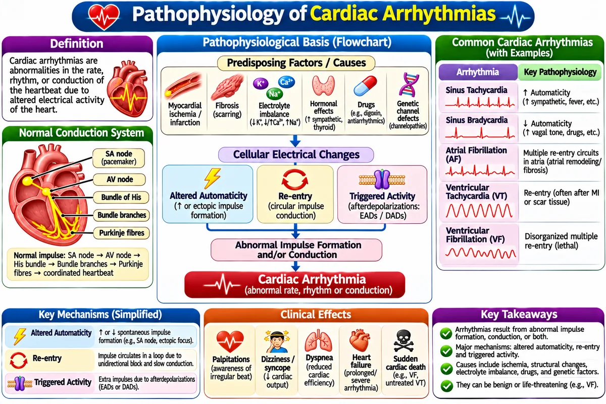
• Sinus Arrhythmias: Sinus Bradycardia (< 60 bpm, seen in athletes or sick sinus syndrome) and Sinus Tachycardia (> 100 bpm, triggered by fever, exercise, or hyperthyroidism).
• Atrial Fibrillation (AF): Chaotic, disorganized atrial electrical depolarization (~400–600 bpm) causing irregularly irregular pulse and loss of atrial kick; stagnation of blood in the left atrial appendage increases stroke risk 5-fold (requires oral anticoagulants like Apixaban or Warfarin).
• Ventricular Fibrillation (VF): Disorganized, chaotic ventricular depolarization with zero coordinated stroke volume and zero cardiac output; causes immediate hemodynamic collapse and sudden cardiac death within minutes unless rapidly converted by DC electrical defibrillation.