Introduction to Human Body, Cell, Tissues, Homeostasis & Cell Injury
Human Anatomy and Physiology (HAP) is defined as the study of the structure and functions of the human body. In this foundational unit, every topic is explained using easy words, simple definitions, clear classifications, and student-friendly diagrams as per the official B. Pharmacy 1st Semester syllabus.
In this topic, you will learn:
1. Definition & Scope of Anatomy & Physiology, Levels of Structural Organisation, 11 Body Systems & Basic Life Processes
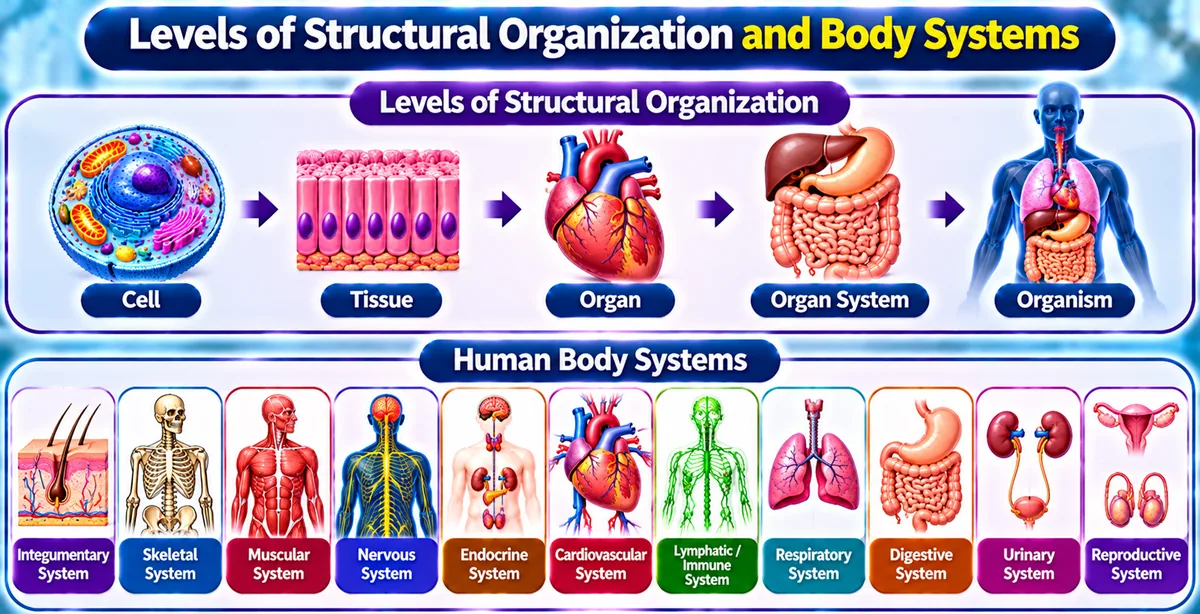
Definition and Scope of Anatomy and Physiology:
- • Human Anatomy & Physiology (HAP): It is defined as it is the study of structure and functions of the human body.
- • Anatomy: It is the branch of science which deal with the study of structure of different organs and body parts of the human body. eg. Histology (study about tissues).
- • Physiology: It is the branch of science which deal with the study of functions of different organs and body parts of the human body. eg. Neurophysiology (study about neurons).
- • Scope of Anatomy & Physiology: It is about what we can do from anatomy and physiology:
- Study of structure and function of body parts.
- Understanding parameters of normal health such as body temperature, blood pressure, pH, and basic nutritional needs.
- Understanding the pathology and mechanisms of diseases.
- Developing surgery techniques and clinical procedures.
- Studying human evolution, growth, and development.
Levels of Structural Organisation:
The human body is a six-level organized structure:
It is the most basic level. Two or more atoms and molecules join together to form cells. eg. Oxygen (O), Carbon (C), Hydrogen (H), DNA, glucose.
It is the basic structural and functional level of the body, i.e. cell. Two or more cells join together to form tissue.
These are the group of cells which works together to perform a particular function. eg. Epithelial, Connective, Muscular, Nervous tissue.
In this, different types of tissue combine together to form organs which do proper functioning of body. eg. Heart, lungs, kidney, stomach.
In this, a group of organs combine together to form a system. eg. Digestive system, Respiratory system, Cardiovascular system.
It is the highest level and a complete body made up with combination of all systems. eg. Human body.
The 11 Body Systems:
A system is a group of organs which combine together to perform proper functioning. There are total (11) systems in the human body:
It coordinates all the actions of the body. It is responsible for all voluntary and involuntary actions and all signalling. organs → Brain, spinal cord, nerves.
It involves respiration and exchange of gases [O₂ ↔ CO₂] between blood and air. organs → Nose, pharynx, larynx, trachea, lungs.
It is responsible for the circulation of blood in the body, that's why it is also known as circulatory system. organs → Heart, blood vessels, blood.
It is responsible for digestion of food and absorbs nutrients from it. organs → Mouth, stomach, intestines, liver, pancreas.
It is responsible for the filtration of blood and also removes waste products as urine. organs → Kidneys, ureters, urinary bladder, urethra.
It is responsible for the production of offspring. organs → Testes, ovaries, fallopian tubes, uterus.
It is also known as exocrine system because it contains skin which provides protection and contains glands. organs → Skin, hair, nails, sweat glands.
It is responsible for the movement of our body organs and skeleton through muscles. organs → Skeletal muscles, tendons.
It contains bones which maintain structure and provide protection to our body organs. organs → Skull, ribs, vertebral column, femur (206 bones).
It is also known as immune system. It defends the body against pathogens that may harm the body. This system consists of a network of lymphatic vessels that carry a clear fluid called lymph. organs → Spleen, thymus, lymph nodes, tonsils.
A system consists of different types of hormones released into the blood which helps in the functioning of the body. organs → Pituitary, thyroid, adrenal glands, pancreas.
Basic Life Processes:
The human body performs different functions for its survival and growth. All living organisms have specific life processes:
- • 1. Metabolism: It is the sum of all chemical processes that occur in the body. It is of two types:
- Catabolism: It is the breakdown of complex chemical substances into simple compounds (releases energy).
- Anabolism: It is the building up of complex chemical substances from small components (uses energy).
- • 2. Responsiveness: It is the ability of the body to detect and respond to changes in the environment. eg. Moving hand away from a hot object, shivering in cold.
- • 3. Movement: It includes the motion of the whole body, individual organs, single cells, and even tiny organelles inside cells.
- • 4. Growth: It is the development of our body and also increase in body size, caused by increase in cell size and number of cells.
- • 5. Differentiation: It is the development of a cell from an unspecialized to a specialized state. eg. Stem cells develop into blood cells, nerve cells, or bone cells.
- • 6. Reproduction: It refers to the formation of new cells for tissue growth and repair, and also produces new offspring.
- • 7. Respiration: It involves the exchange of O₂ and CO₂ between body cells and the external environment.
- • 8. Digestion: It involves the breakdown of food molecules into small units and absorption of nutrients into the blood.
- • 9. Excretion: It is the process of removal of metabolic waste products from the body. eg. Urination (removal of urea and water).
2. Homeostasis and Feedback Systems
Homeostasis (In Simple Words):
Homeostasis is derived from two Greek words: Homeo (meaning same or constant) + stasis (meaning standing still or state). It means staying the same.
It is defined as a condition when our internal environment remains constant with respect to changes in the external environment. It is a condition that may vary but remains relatively constant within normal limits.
- • It is also known as: The dynamic equilibrium or internal biological balance of the body.
- • It is used for: Maintaining parameters of normal health: core body temperature stays at 37°C, arterial blood pH stays at 7.35–7.45, and fasting blood glucose stays at 70–100 mg/dL.
- • Homeostasis Control Mechanism: All body organs coordinate with each other to maintain homeostasis. This coordination is mainly controlled by the Neuroendocrine System (Nervous system + Endocrine system).
- • eg. Body Temperature: When it is cold outside, your body shivers to produce heat; when it is hot outside, your skin sweats to cool down, keeping your body at 37°C constant.
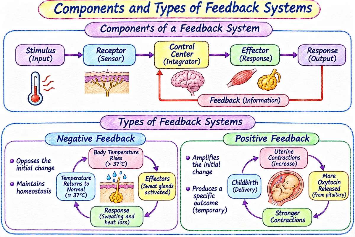
The 3 Components of a Homeostatic Feedback System:
It has three components that work together to maintain internal balance:
Types of Feedback Systems:
Feedback systems are of two types:
When anything increases in our internal environment (body), then this system tries to bring it back into normal condition by decreasing it. Similarly, when anything decreases, it brings it back by increasing it.
• Body Temperature: When temperature rises, skin receptors signal the hypothalamus → stimulates sweat glands → sweating cools body down to 37°C.
• Blood Glucose: When blood sugar rises after food, pancreas releases insulin → cells absorb glucose → blood sugar returns to normal.
When anything changes in the internal environment, the positive feedback system strengthens and amplifies that change until a specific event is completed.
• Childbirth (Parturition): Contraction of uterus pushes baby against cervix → signals release of oxytocin hormone → oxytocin increases uterine contraction → cycle repeats until childbirth is complete.
• Blood Clotting: Damaged vessel activates platelets → platelets release chemicals that attract more platelets → forms a plug to stop bleeding.
3. Basic Anatomical Terminologies (Directional Terms, Planes & Cavities)
Simple Concept:
Anatomical terminology is defined as the standardized language used by doctors, pharmacists, and scientists to describe the exact position, orientation, planes, and cavities of body parts. It is divided into three groups: 1) Directional terms, 2) Sectional planes, and 3) Body cavities.
1. Directional Terms:
| Directional Term | Simple Meaning | Clear Example |
|---|---|---|
| Superior (Cranial) | Upper part, towards the head | The forehead is superior to the nose. |
| Inferior (Caudal) | Lower part, away from the head | The stomach is inferior to the lungs. |
| Anterior (Ventral) | Front part of the body | The sternum (breastbone) is anterior to the heart. |
| Posterior (Dorsal) | Back side of the body | The esophagus is posterior to the trachea. |
| Medial | Nearer to the midline of the body | The ulna is on the medial side of the forearm. |
| Lateral | Farther from the midline of the body | The lungs are lateral to the heart. |
| Proximal | Nearer to the point of attachment of a limb | The humerus (arm bone) is proximal to the radius. |
| Distal | Farther from the point of attachment of a limb | The phalanges (fingers) are distal to the wrist. |
- • Sagittal Plane: It divides the body vertically into right and left parts. (Midsagittal divides into equal right and left halves).
- • Coronal / Frontal Plane: It divides the body vertically into anterior (front) and posterior (back) parts.
- • Transverse Plane: It is a horizontal plane parallel to the ground that passes through the body at 90° angle and divides the body into superior (upper) and inferior (lower) parts.
- • Dorsal Body Cavity: Located near the back; it contains the Cranial cavity (protects the brain) and Vertebral canal (protects spinal cord).
- • Thoracic Cavity: Chest cavity enclosed by ribs; it contains the heart (in pericardial cavity) and lungs (in pleural cavities).
- • Abdominal & Pelvic Cavity: It contains the stomach, intestines, liver, spleen, kidneys (abdominal), and urinary bladder, internal reproductive organs (pelvic).
4. Cell Structure, Membrane Transport, and Intercellular Signalling
Simple Definition:
The Cell is defined as the basic structural and functional unit of all living organisms. Every human cell consists of three main parts: 1) Plasma membrane, 2) Cytoplasm containing organelles, and 3) Nucleus.
The Plasma Membrane (Fluid Mosaic Model):
It is also known as cell membrane. It consists of a lipid bilayer made of phospholipids with cholesterol and proteins floating in it like a mosaic:
- Phospholipids: Have hydrophilic (water-loving) heads and hydrophobic (water-fearing) tails.
- Proteins: Act as ion channels, carrier transporters, receptors for hormones, and cell markers.
- Selective Permeability: It allows small, non-polar molecules (O₂, CO₂) to pass easily, but controls large or charged molecules (glucose, Na⁺).
Key Organelle Functions:
- • Mitochondria: Also known as the powerhouse of the cell. It is responsible for aerobic cellular respiration and generates ATP energy.
- • Ribosomes: Responsible for protein synthesis.
- • Endoplasmic Reticulum (ER): Rough ER (with ribosomes) helps in synthesizing proteins; Smooth ER helps in synthesizing lipids and detoxifying drugs.
- • Golgi Apparatus: Responsible for sorting, packaging, and secretion of proteins into vesicles.
- • Lysosomes: Also known as suicidal bags of the cell. Contains digestive enzymes that break down waste and damaged organelles.
- • Nucleus: Contains DNA genetic material and controls all cell activities.
Membrane Transport Mechanisms: Passive vs. Active Transport
Transport across cell membrane is of two main types:
| Transport Type | Energy (ATP) Needed? | Direction of Movement | Clear Example |
|---|---|---|---|
| Simple Diffusion | NO (Passive) | Down concentration gradient (High → Low) | Oxygen and CO₂ moving across alveolar capillary membrane. |
| Facilitated Diffusion | NO (Passive) | Down gradient via transport protein channels | Glucose transport into cells via GLUT transporters. |
| Osmosis | NO (Passive) | Water moves from low solute to high solute area | Movement of water across red blood cell membrane in isotonic solution. |
| Primary Active Transport | YES (Direct ATP) | Against gradient (Low → High) | Na⁺/K⁺ ATPase Pump: Pumps 3 Na⁺ out and 2 K⁺ in per ATP. |
| Endocytosis / Exocytosis | YES (ATP required) | Vesicle movement across membrane | Phagocytosis by macrophages (cell eating) and secretion of insulin. |
How Cells Talk to Each Other (The 4 Communication Forms):
Cells coordinate with each other through four primary signalling methods:
A cell releases a chemical messenger that binds to receptors on its own surface. eg. T-lymphocytes release Interleukin-2 to stimulate their own proliferation.
A cell releases messengers that act on nearby neighbouring target cells. eg. Histamine released by mast cells during inflammation acts on nearby blood vessels.
Endocrine glands release hormones into the bloodstream to reach distant target tissues throughout the body. eg. Thyroid hormone, Insulin.
Neurons release neurotransmitters across a tiny synaptic gap (~20 nm) directly to target cells. eg. Acetylcholine at neuromuscular junction.
5. The Four Basic Tissue Classes
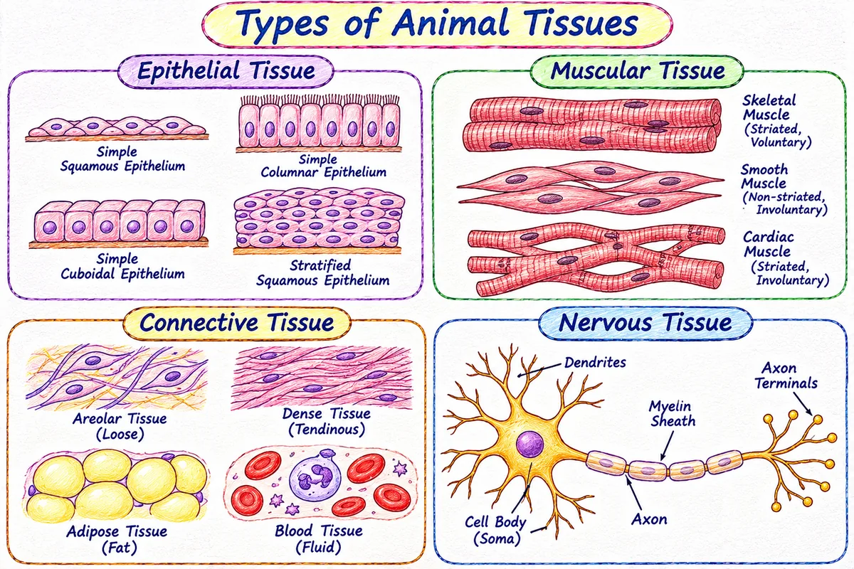
The 4 Primary Tissues (Explained in Simple Terms):
A tissue is defined as a group of similar cells which works together to perform a particular function. There are total (4) primary types of tissues in the human body: 1) Epithelial tissue (covers and protects), 2) Connective tissue (supports and connects), 3) Muscular tissue (produces movement), and 4) Nervous tissue (controls and signals).
1. Epithelial Tissue
It is defined as a continuous sheet of closely packed cells with very little extracellular matrix, resting on a basement membrane. It covers body surfaces, lines hollow organs, cavities, and ducts, and forms glands.
- Functions: It is responsible for protection, absorption, secretion, and filtration.
- Types: Simple Epithelium (single layer, used for absorption/filtration: simple squamous in lung alveoli, simple cuboidal in kidney tubules, simple columnar in intestine) and Stratified Epithelium (multiple layers, used for protection against friction: stratified squamous in skin epidermis).
2. Connective Tissue
It is the most abundant and widely distributed tissue in the body. It consists of cells separated by a large amount of extracellular matrix (fibres + ground substance).
- Functions: It protects, supports, binds organs together, stores energy as fat, and provides immunity.
- Types: Loose CT (areolar, adipose), Dense CT (tendons, ligaments), Cartilage (hyaline, elastic, fibrocartilage), Bone tissue (compact, spongy), and Liquid CT (blood and lymph).
3. Muscular Tissue
It consists of elongated muscle cells (muscle fibres) specialized for contraction and generating physical force. It is of three types:
- Skeletal Muscle: Striated, cylindrical, multinucleated, voluntary control. Attached to bones for movement.
- Cardiac Muscle: Striated, branched, involuntary control, intercalated discs. Forms wall of the heart to pump blood.
- Smooth Muscle: Non-striated, spindle-shaped, single nucleus, involuntary control. Located in walls of hollow internal organs (stomach, blood vessels).
4. Nervous Tissue
It detects changes in internal and external environment and generates electrical nerve impulses to coordinate body functions.
- Neurons: Structural and functional units of nervous system. Consist of cell body (soma), dendrites (receive impulses), and axon (transmits impulses).
- Neuroglia (Glial Cells): Supporting cells that protect, nourish, and insulate neurons (astrocytes, oligodendrocytes, microglia, Schwann cells).
6. Pathogenesis of Cell Injury and Cellular Adaptive Changes
Simple Definition:
Cell Injury is defined as a condition when a cell is exposed to harmful stress or noxious agents that exceed its normal ability to adapt, leading to damage to cell structure and its functions. It is of two types: 1) Reversible cell injury (cell can recover if stress is removed), and 2) Irreversible cell injury (severe damage leads to cell death).
Causes of Cell Injury (Etiology):
Cell injury can occur due to several causes:
Lack of oxygen (hypoxia) or reduced blood flow (ischemia) is the most common cause of cell injury. Inhibits ATP production.
Mechanical trauma, extremes of temperature (burns, frostbite), radiation, and electric shock.
Poisons (cyanide, arsenic), alcohol, environmental pollutants, and high drug overdoses.
Pathogenic bacteria, viruses, fungi, and parasites that invade cells and release toxins.
Allergic hypersensitivity reactions and autoimmune diseases where the immune system attacks own body cells.
Gene mutations (sickle cell anaemia) and nutritional deficiencies (protein malnutrition, vitamin lack).
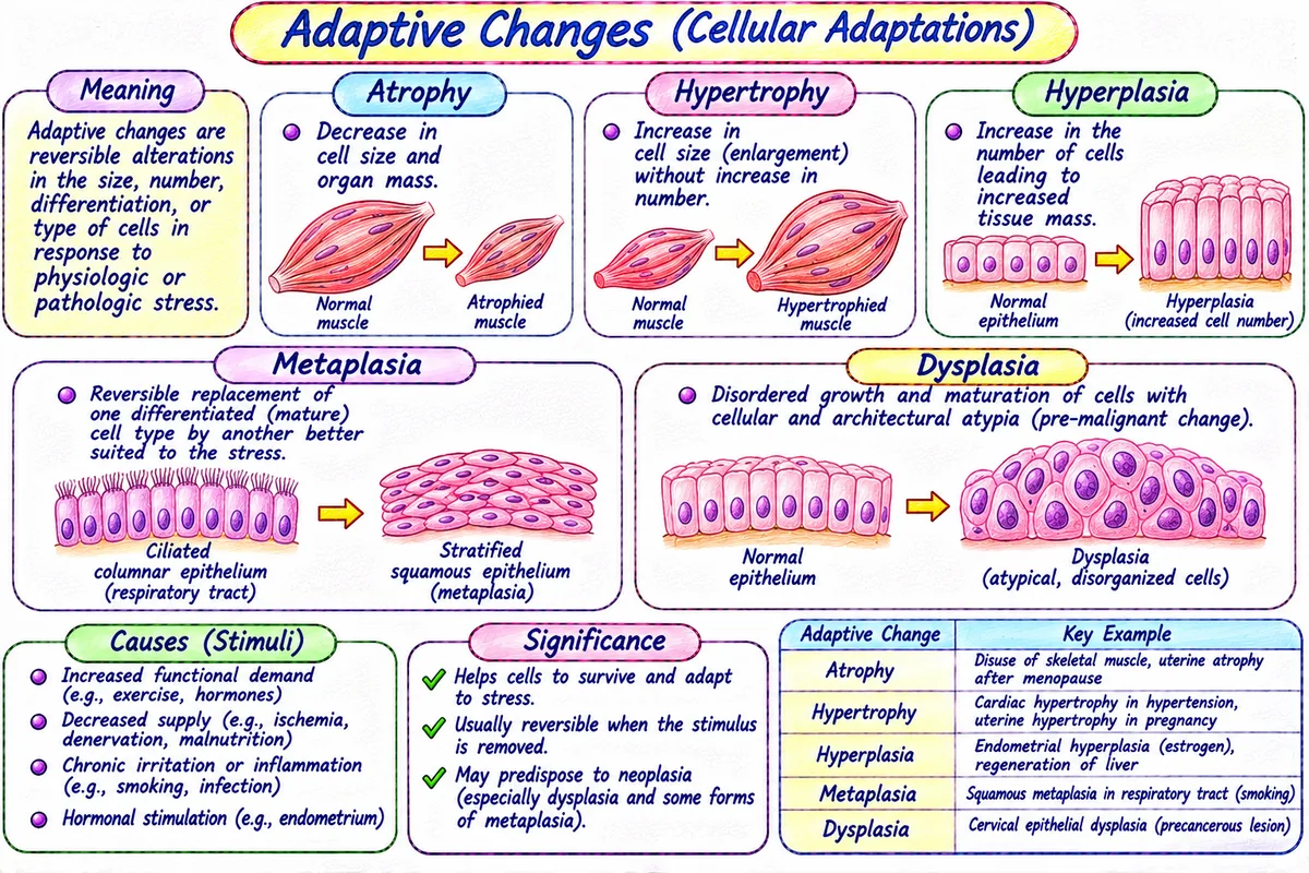
Cellular Adaptive Changes (Atrophy, Hypertrophy, Hyperplasia, Metaplasia, Dysplasia):
When cells face persistent stress, they adapt their size, number, or phenotype to survive. It is of five types:
It is defined as: Decrease in cell size and shrinkage of organ due to loss of cell substance. eg. Shrinkage of arm muscles immobilized in plaster cast.
It is defined as: Increase in the size of cells resulting in enlarged organ size without cell division. eg. Growth of skeletal muscle in weightlifters; left ventricular hypertrophy in hypertension.
It is defined as: Increase in the number of cells in an organ or tissue. eg. Enlargement of breast tissue and uterus during pregnancy.
It is defined as: Reversible transformation of one adult cell type into another adult cell type. eg. In chronic cigarette smokers, ciliated columnar respiratory epithelium transforms into tough stratified squamous epithelium.
It is defined as: Abnormal, disordered growth and maturation of cells with loss of architectural arrangement. It is also known as pre-cancerous change. eg. Cervical dysplasia detected by Pap smear.
7. Cell Death: Necrosis vs. Apoptosis
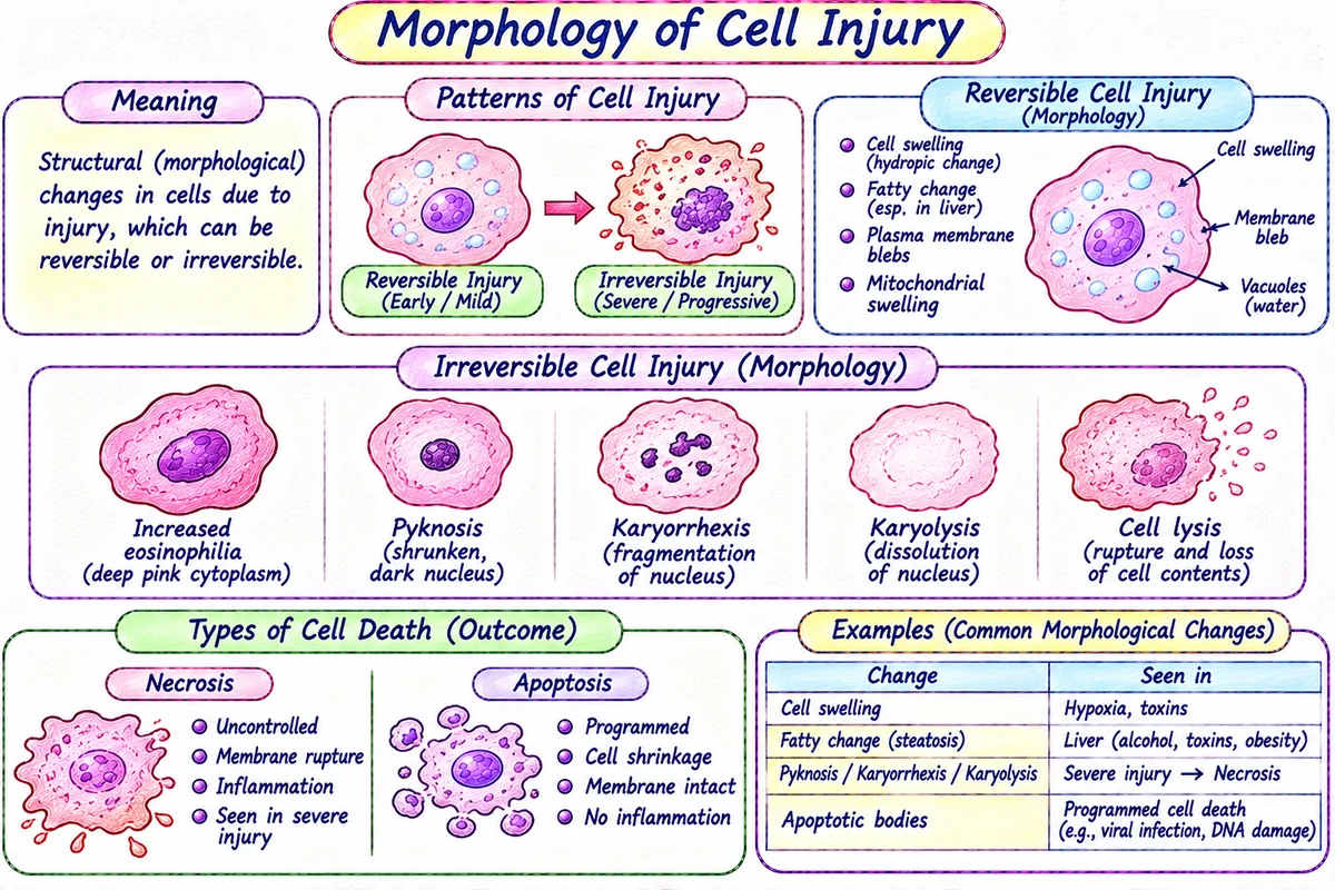
Cell Death (In Simple Words: Accidental Death vs. Programmed Suicide):
When cell injury becomes irreversible, cell death occurs. There are two major types of cell death:
- • Necrosis ("Cell Murder"): It is defined as accidental, unprogrammed cell death caused by severe injury, infection, or lack of blood supply. Cells swell up, burst, and spill enzymes into surrounding tissue, triggering severe inflammation.
- • Apoptosis ("Cell Suicide"): It is defined as programmed, clean cell death where old, unwanted, or damaged cells pack themselves neatly and self-destruct without causing any inflammation.
| Feature | Necrosis ("Cell Murder")• It is also known as: Accidental cell death.• It is caused by: Severe external trauma, hypoxia, or toxins. | Apoptosis ("Cell Suicide")• It is also known as: Programmed cell death (PCD).• It is used for: Clean removal of unwanted or damaged cells. |
|---|---|---|
| Nature of Process | Always pathological (disease-caused), involuntary and passive. | Mostly physiological (normal tissue turnover), tightly gene-regulated and active (requires ATP). |
| Cell Size & Shape | Cell swells up (cellular edema). | Cell shrinks and rounds up. |
| Plasma Membrane | Ruptures and breaks down; cell contents leak out. | Remains intact; forms small membrane-bound apoptotic bodies. |
| Inflammation | YES → Causes intense local inflammation and tissue damage. | NO → Zero inflammation; apoptotic bodies are quietly swallowed by macrophages. |
| Clear Examples | Myocardial infarction (heart muscle death during heart attack), gangrene in limb. | Separation of webbed fingers in human embryo during development; monthly shedding of endometrial lining during menstruation. |