Peripheral Nervous System & Special Senses
The peripheral nervous system connects our brain and spinal cord to every muscle, organ, and sensory receptor in the body. In this unit, we explain the autonomic nervous system (sympathetic vs parasympathetic), the 12 pairs of cranial nerves and 31 pairs of spinal nerves, the anatomy and physiology of special senses (eye, ear, nose, tongue), and common sensory disorders (glaucoma, cataract, myopia, otitis, vertigo, and anosmia).
In this topic, you will learn:
1. Classification of PNS: Sympathetic vs. Parasympathetic Nervous System
Simple Definition:
The Peripheral Nervous System (PNS) is defined as all nervous tissue existing outside the Central Nervous System (brain and spinal cord). It is functionally subdivided into the Somatic Nervous System (SNS) (voluntary control of skeletal muscles) and the Autonomic Nervous System (ANS) (involuntary regulation of smooth muscles, cardiac muscle, and glands).
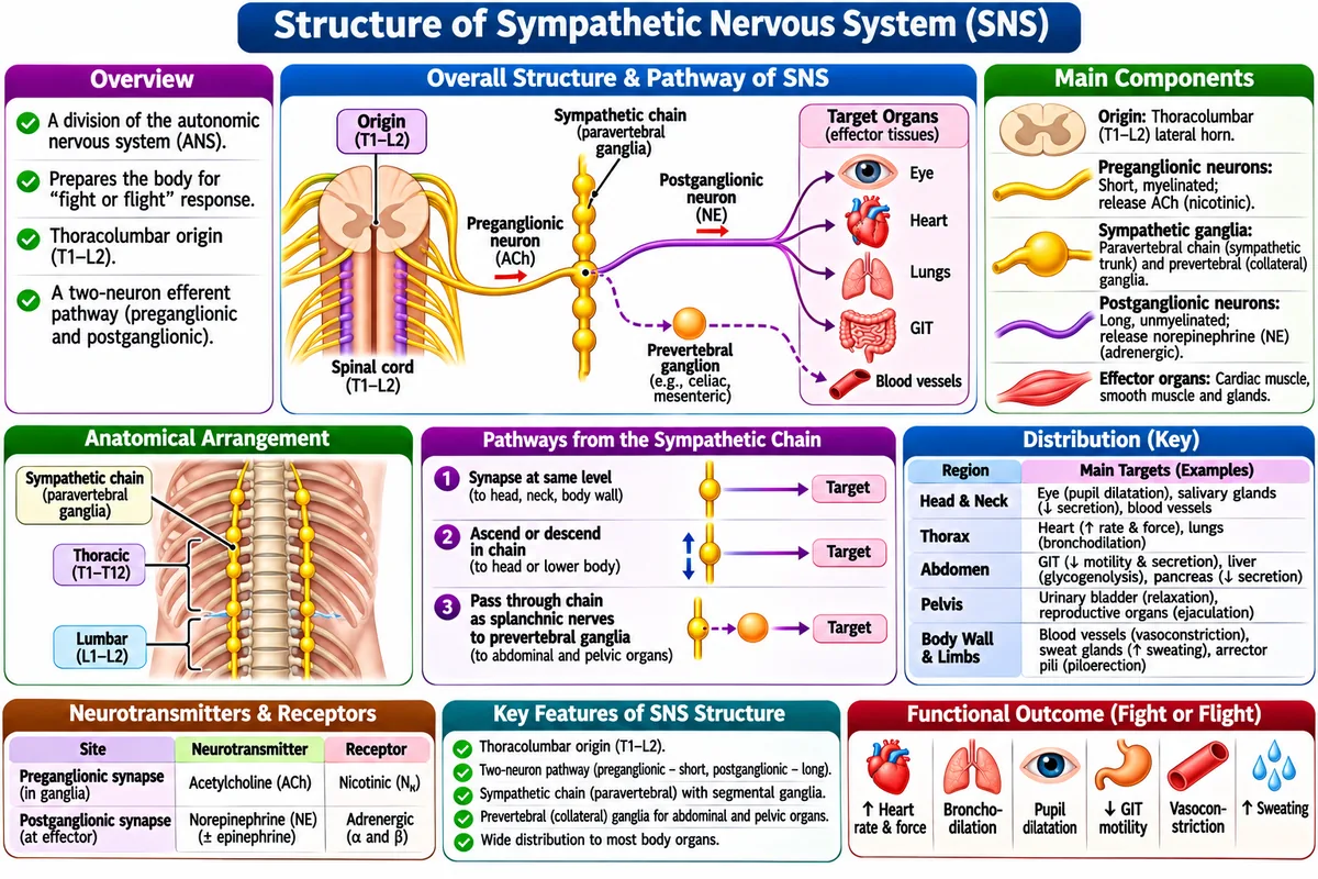
1. Sympathetic Nervous System ("Fight-or-Flight")
It is also known as: Thoracolumbar Outflow (spinal segments T1 through L2). It is used for: mobilizing stored metabolic energy and accelerating cardiopulmonary activity during physical exertion, stress, emergencies, or physiological shock.
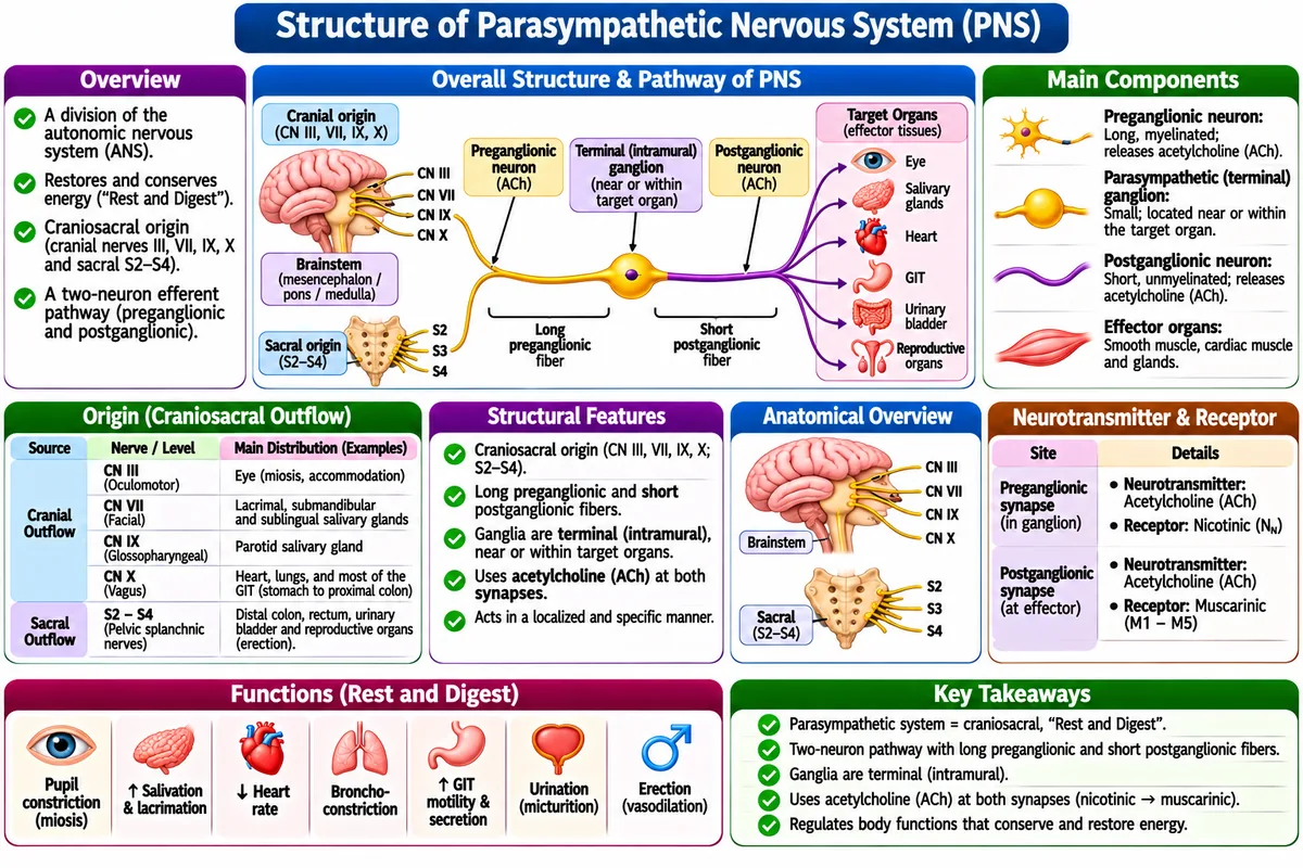
2. Parasympathetic Nervous System ("Rest-and-Digest")
It is also known as: Craniosacral Outflow (cranial nerves III, VII, IX, X and sacral segments S2 through S4). It is used for: conserving energy, lowering heart rate, promoting peristalsis and enzymatic digestion, and restoring baseline homeostasis.
3. Comparison: Sympathetic vs. Parasympathetic Division
| Feature / Target Organ | Sympathetic System ("Fight-or-Flight")• It is also known as: Thoracolumbar outflow.• It is used for: Mobilizing energy during emergency physical threats. | Parasympathetic System ("Rest-and-Digest")• It is also known as: Craniosacral outflow.• It is used for: Conserving bodily energy and promoting digestion and rest. |
|---|---|---|
| Anatomical Outflow | Thoracolumbar Outflow (T1 to L2 spinal segments) | Craniosacral Outflow (Cranial nerves III, VII, IX, X + Sacral S2–S4) |
| Neurotransmitters | Preganglionic = ACh; Postganglionic = Noradrenaline (Norepinephrine) | Preganglionic = ACh; Postganglionic = Acetylcholine (ACh) |
| Heart (Cardiac Muscle) | Increases heart rate and force of contraction (β₁ receptors). | Decreases heart rate (bradycardia via M₂ muscarinic receptors). |
| Lungs (Bronchioles) | Bronchodilation (opens airways via β₂ receptors). | Bronchoconstriction and increased mucus secretion (M₃ receptors). |
| Eye (Pupil) | Mydriasis (pupil dilation via α₁ to see threats in distance). | Miosis (pupil constriction via M₃ for close near vision). |
| GI Tract & Digestion | Inhibits peristalsis and shuts sphincters. | Stimulates gastric acid, saliva secretion, and bowel peristalsis. |
2. Spinal Nerves (31 Pairs) and Cranial Nerves (12 Pairs)
The 31 Pairs of Spinal Nerves:
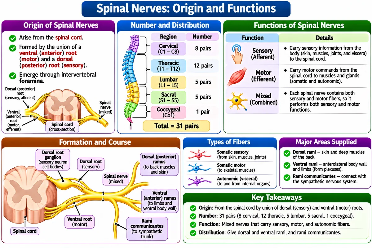
All spinal nerves are mixed nerves (carrying both sensory afferent dorsal roots and motor efferent ventral roots):
The 12 Cranial Nerves:
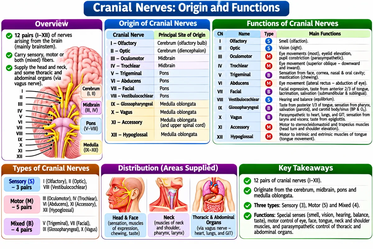
Memory Mnemonic: "On Old Olympus' Towering Top, A Finn And German Viewed Some Hops"
| No. | Cranial Nerve Name | Type (Sensory/Motor/Both) | Primary Physiological Function |
|---|---|---|---|
| I | Olfactory | Sensory | Sense of smell (olfaction). |
| II | Optic | Sensory | Vision (transmits image signals from retina to occipital lobe). |
| III | Oculomotor | Motor | Controls 4 extrinsic eye muscles + pupillary constriction and lens focusing. |
| IV | Trochlear | Motor | Superior oblique eye muscle (moves eye inferolaterally). |
| V | Trigeminal (Largest) | Both | Sensory to face, teeth, cornea; Motor to muscles of mastication (chewing). |
| VI | Abducens | Motor | Lateral rectus eye muscle (abducts eye laterally). |
| VII | Facial | Both | Taste anterior 2/3 tongue; Motor to facial expression muscles (paralysis = Bell's Palsy). |
| VIII | Vestibulocochlear | Sensory | Hearing (Cochlear branch) and dynamic/static equilibrium (Vestibular branch). |
| IX | Glossopharyngeal | Both | Taste posterior 1/3 tongue; swallowing; salivation from parotid gland. |
| X | Vagus ("Wanderer") | Both | Controls 75% of parasympathetic outflow: slows heart, stimulates digestion in stomach, intestines. |
| XI | Accessory | Motor | Sternocleidomastoid and Trapezius muscles (shrugging shoulders, turning head). |
| XII | Hypoglossal | Motor | Intrinsic and extrinsic muscles of tongue (speech and swallowing). |
3. Structure and Functions of Special Senses: Eye, Ear, Nose and Tongue
A. The Human Eye (Organ of Vision)
The eye is a complex sensory organ situated within the orbital cavity, specialized for photoreception and image formation. The eyeball consists of three distinct anatomical coats (tunics) surrounding fluid-filled optical chambers:
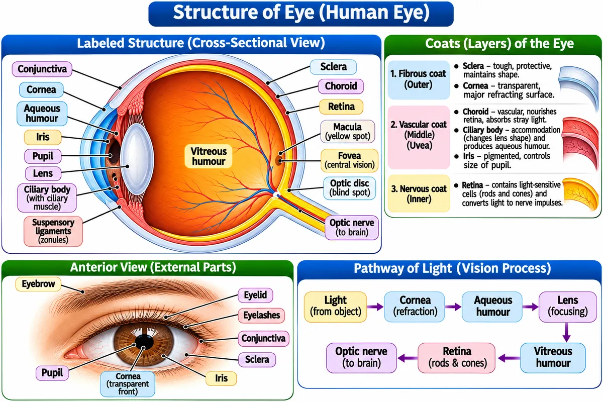
• 1. Fibrous Tunic (Outer): Anterior transparent Cornea (responsible for ~70% of total light refraction) and posterior white, fibrous Sclera (maintains eyeball shape and anchors extraocular muscles).
• 2. Vascular Tunic / Uvea (Middle): Heavily pigmented Choroid (nourishes retina and absorbs stray light), Ciliary Body (ciliary processes secrete aqueous humor; ciliary muscles control lens accommodation), and Iris (colored contractile diaphragm with sphincter and dilator pupillae regulating light entry through pupil).
• 3. Neural Tunic / Retina (Inner): The sensory receptor layer containing ~120 million Rods (rhodopsin pigment; specialized for low-light scotopic night vision and peripheral movement) and ~6 million Cones (photopsin pigments; specialized for high-acuity color photopic vision, concentrated at the Fovea Centralis within the macula lutea).
B. The Human Ear (Organ of Hearing and Equilibrium)
The ear functions as a dual-modality sensory apparatus transducing acoustic vibrational waves into auditory perceptions, while simultaneously monitoring head position and kinetic acceleration for balance. It is divided into three chambers:
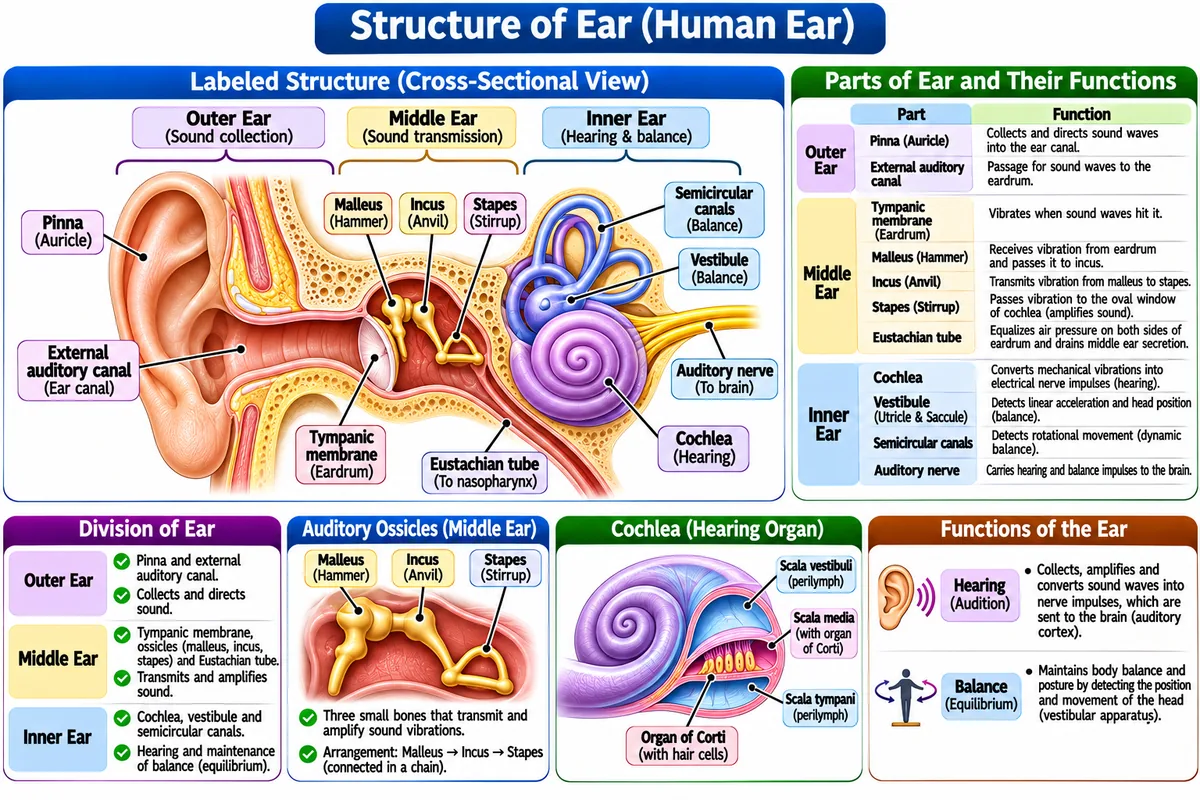
• 1. External Ear: Auricle (pinna) and external acoustic meatus lined with ceruminous glands; directs sound waves onto the Tympanic Membrane.
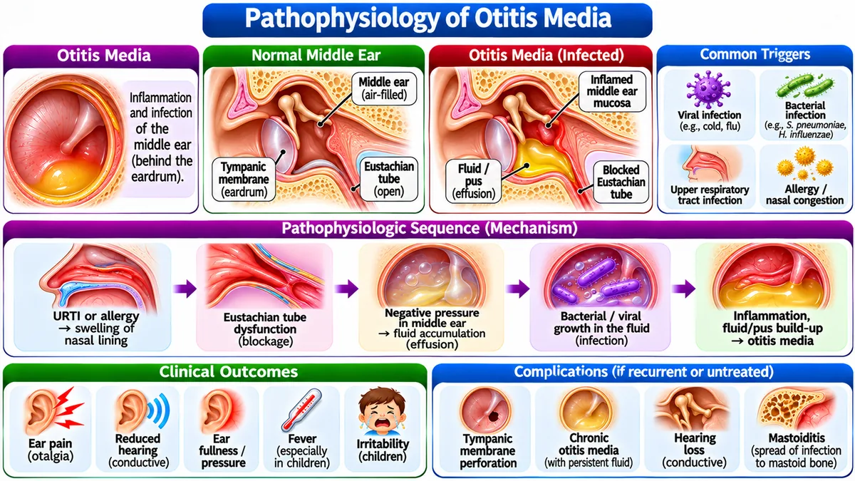
• 2. Middle Ear (Tympanic Cavity): Air-filled petrous temporal bone chamber housing 3 auditory ossicles: Malleus → Incus → Stapes. These bones mechanically amplify acoustic sound energy ~20-fold onto the oval window. Connected to the nasopharynx via the Eustachian tube to equalize middle ear atmospheric pressure.
• 3. Inner Ear (Bony & Membranous Labyrinth): Houses the snail-shaped Cochlea containing the Organ of Corti where sensory hair cells convert fluid pressure waves into nerve impulses (Cranial Nerve VIII); and the Vestibular Apparatus (3 Semicircular Canals for rotational acceleration; Utricle and Saccule with otolith organs for linear acceleration and gravitational equilibrium).
C. The Nose (Organ of Olfaction)
Olfaction (sense of smell) is a direct chemical sensation mediated by the olfactory neuroepithelium occupying the roof of the nasal cavity across the superior concha and nasal septum.
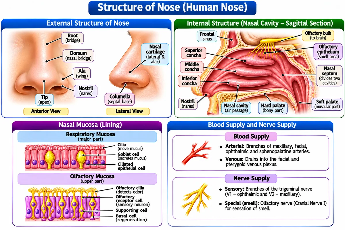
• Olfactory Receptors: Composed of ~10–100 million specialized bipolar receptor neurons with apical non-motile cilia embedded in mucus secreted by Bowman's glands.
• Signal Transduction: Volatile airborne odorants dissolve in mucus and bind specific G-protein coupled olfactory receptors (Golf), activating Adenylyl Cyclase → cAMP generation → Na⁺/Ca²⁺ channel opening → depolarization.
• Direct Limbic Transmission: Unmyelinated axons traverse the cribriform plate of the ethmoid bone to synapse in the Olfactory Bulb (Cranial Nerve I). Olfactory signals project directly to the primary olfactory cortex and limbic system (amygdala/hippocampus) without thalamic relay, explaining why smells trigger vivid emotional memories.
D. The Tongue (Organ of Gustation)
Gustation (taste) is mediated by ~10,000 taste buds distributed across specialized papillae on the dorsal mucosal surface of the tongue, soft palate, and epiglottis.
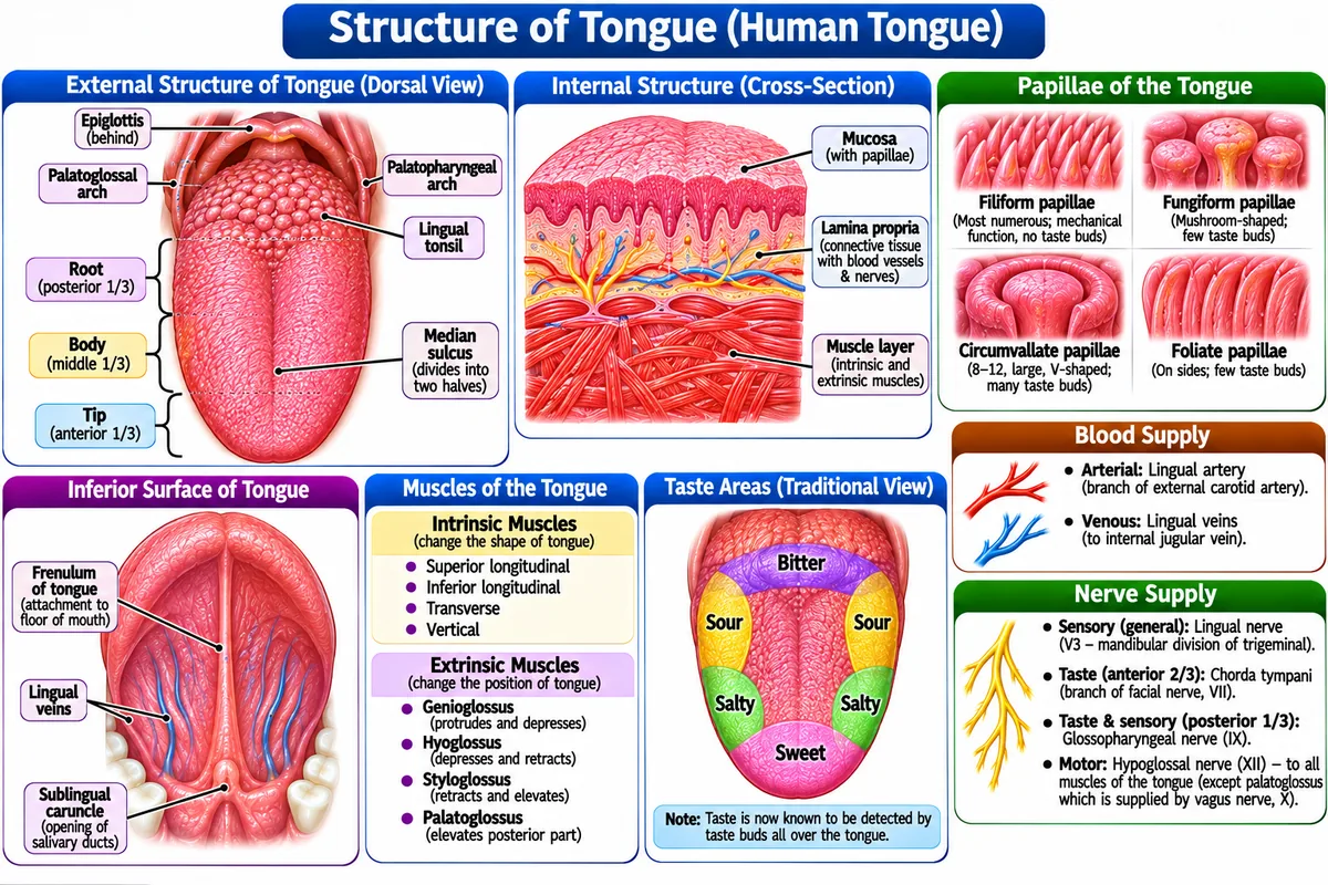
• Papillary Types: Vallate (Circumvallate) forming a V-shape at posterior tongue (contain highest concentration of taste buds); Fungiform scattered across anterior two-thirds; Foliate along lateral folds; and Filiform (tactile keratinized spikes lacking taste buds).
• 5 Primary Taste Modalities: Sweet (sugars/sweeteners via T1R2/T1R3 GPCRs), Salty (direct Na⁺ influx through ENaC channels), Sour (H⁺ protons blocking K⁺ channels), Bitter (toxins/alkaloids via ~25 T2R GPCRs with lowest detection threshold), and Umami (L-glutamate/monosodium glutamate via T1R1/T1R3 GPCRs).
• Innervation: Anterior 2/3 carries taste via Chorda Tympani (Cranial Nerve VII); posterior 1/3 via Glossopharyngeal (Cranial Nerve IX); and base/epiglottis via Vagus (Cranial Nerve X).
4. Pathophysiology of Special Sense Disorders
1. Glaucoma
Glaucoma is defined as a progressive optic neuropathy associated with elevated intraocular pressure (IOP > 21 mmHg) that leads to retinal ganglion cell death.
• It is also known as: The sneak thief of sight.
• It is used for: Diagnosing optical drainage blockage and guiding pressure-lowering eye drops (e.g., Timolol, Latanoprost).
• In this mechanism, we: See impaired drainage of aqueous humor through the trabecular meshwork cause mechanical compression of the optic nerve head.
Pathophysiology: Glaucoma is a progressive optic neuropathy characterized by accelerated retinal ganglion cell death and optic nerve head cupping. The primary risk factor is pathological elevation of Intraocular Pressure (IOP > 21 mmHg; normal = 10–21 mmHg) resulting from an imbalance between aqueous humor secretion by the ciliary epithelium and drainage through the trabecular meshwork and Canal of Schlemm.
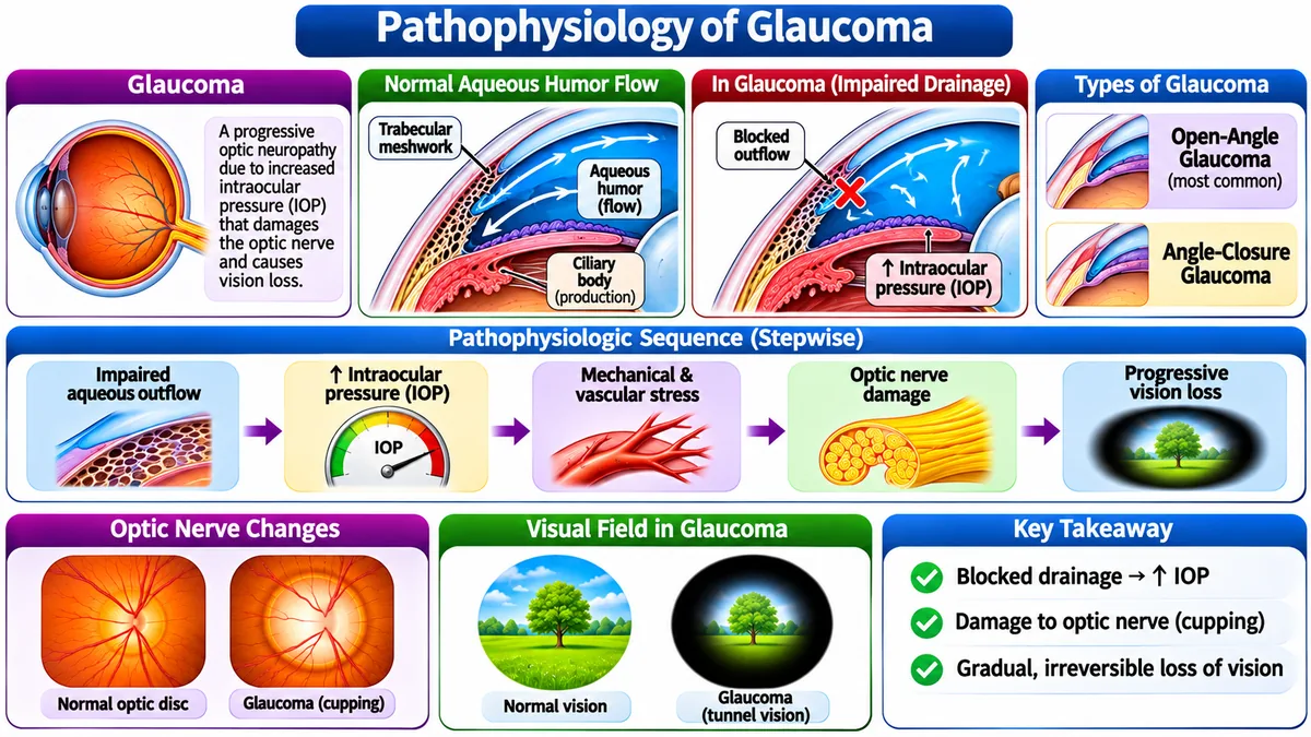
• Primary Open-Angle Glaucoma (POAG): Most common; microscopic degenerative blockage within trabecular meshwork causes insidious, painless, progressive peripheral visual field loss ("tunnel vision").
• Closed-Angle (Angle-Closure) Glaucoma: Iris physically blocks trabecular meshwork angle; rapid severe IOP spike (> 50 mmHg) with excruciating ocular pain, steamy cornea, halo vision, and vomiting; a medical ophthalmic emergency.
• Pharmacotherapy: Prostaglandin analogues (Latanoprost: increases uveoscleral outflow), β-blockers (Timolol: reduces aqueous secretion), and Carbonic Anhydrase inhibitors (Dorzolamide).
2. Cataract
Cataract is defined as the progressive opacification and clouding of the normally crystalline natural lens of the eye.
• It is also known as: Crystalline lens opacification.
• It is used for: Explaining painless, progressive loss of visual acuity and sensitivity to light glare.
• For example: Corrected permanently by ultrasonic phacoemulsification with artificial intraocular lens implantation.
Pathophysiology: Cataract denotes the progressive, painless clouding and opacification of the normally crystalline, transparent biological eye lens. The human lens consists of tightly packed crystallin proteins that maintain optical clarity. With advancing age, systemic diseases (diabetes mellitus), or chronic UV exposure, free radical oxidative stress causes crystallin denaturation, cross-linking, and aggregation into insoluble opacities.
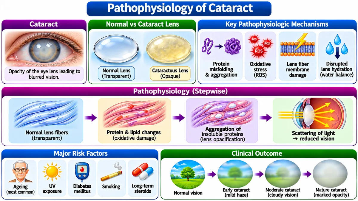
• Clinical Signs: Progressive painless blurring of vision, reduced contrast sensitivity, glare/halos around headlights at night, and fading of color vibrancy.
• Definitive Management: Surgical extraction of cataractous lens via ultrasound phacoemulsification followed by implantation of an artificial Intraocular Lens (IOL).
3. Myopia (Nearsightedness)
Pathophysiology: Myopia is an axial refractive error where the refractive power of the cornea and lens exceeds the anteroposterior axial length of the eyeball. Parallel incoming light rays converge and focus to a sharp point in front of the retina rather than precisely on the photoreceptor layer. As a consequence, near objects appear distinct and crisp, while distant objects appear blurred and unfocused.
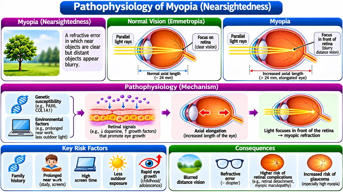
• Comparison with Other Refractive Errors: In Hypermetropia (farsightedness), the eye is axially too short and rays focus behind the retina (corrected with convex lenses); in Astigmatism, irregular corneal curvature creates multiple focal points (corrected with cylindrical lenses).
• Correction: Optical neutralization using Concave (diverging / negative diopter) lenses to diverge incoming light rays before entering the cornea, shifting focal point posteriorly onto the fovea centralis; or refractive corneal surgery (LASIK).
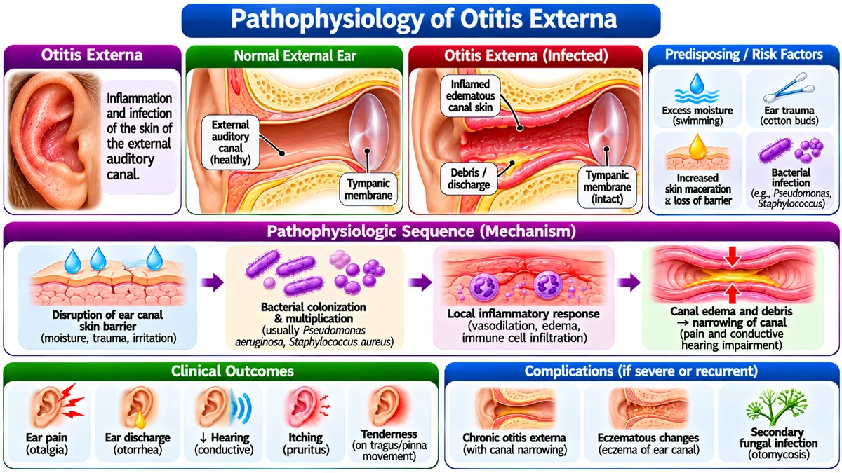
4A. Otitis Externa ("Swimmer's Ear")
Infection of the skin lining the external auditory canal:
4B. Acute Otitis Media (AOM)
Bacterial infection of the mucosal lining of the middle ear cavity behind the intact tympanic membrane:
5. Vertigo & Anosmia
Neurological and vestibular disorders disrupting spatial orientation and chemical sensation:
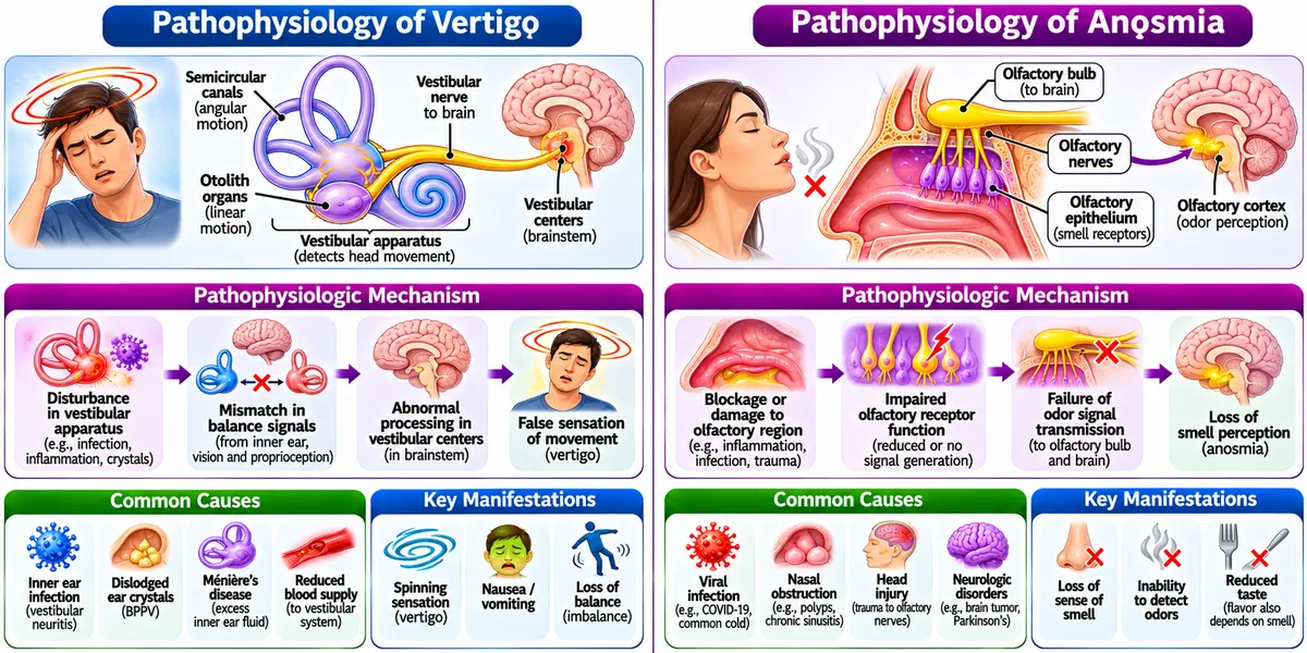
- Peripheral Vertigo: Pathology in semicircular canals or vestibular nerve (CN VIII). Most commonly Benign Paroxysmal Positional Vertigo (BPPV) due to dislodged canalith calcium carbonate otoconia floating in the posterior semicircular canal (treated by Epley repositioning maneuver); or Meniere's Disease (endolymphatic hydrops triad: episodic vertigo, tinnitus, sensorineural hearing loss).
- Central Vertigo: Pathology in brainstem vestibular nuclei or cerebellum (stroke, multiple sclerosis).