Skin, Skeletal System, Muscle Contraction & Joints
Our skin acts as an impenetrable physical barrier, while the skeletal framework of 206 bones and synovial joints allows us to stand upright and move under the power of skeletal muscle fibers. In this unit, we examine skin histology, wound healing, axial and appendicular skeletal bones, the sliding filament theory of muscle contraction, neuromuscular junctions, and joint diseases (rheumatoid arthritis, osteoporosis, and gout).
In this topic, you will learn:
1. Structure and Functions of Skin and Common Skin Disorders
Simple Definition:
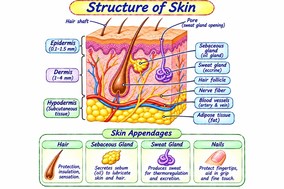
The Integumentary System is defined as the external organ system comprising the skin (the largest organ of the human body, weighing ~4–5 kg) and its accessory appendages (hair, nails, sweat glands, and sebaceous glands).
• It is also known as: The avascular keratinized outer shield.
• It is used for: Protecting underlying tissues from mechanical abrasion, water evaporation, bacteria, and ultraviolet (UV) radiation.
Keratinized stratified squamous epithelium. Formed of 5 microscopic layers (from superficial to deep):
1. Stratum Corneum: Dead flat keratin scales.
2. Stratum Lucidum: Clear layer (palms & soles only).
3. Stratum Granulosum: Keratohyalin granules.
4. Stratum Spinosum: Prickle cell layer.
5. Stratum Basale: Actively dividing stem cells & Melanocytes (produce melanin UV pigment).
• It is also known as: The vascular living core of the skin.
• It is used for: Providing tensile strength and elasticity, nourishing the epidermis, and housing touch and temperature nerve receptors.
Tough, flexible connective tissue layer rich in collagen and elastin fibers:
- Papillary Layer: Dermal papillae forming unique fingerprint ridges.
- Reticular Layer: Houses blood vessels, nerve endings (Meissner's & Pacinian corpuscles for touch and pressure), hair follicles, and sweat glands.
• It is also known as: Subcutaneous tissue (Subcutis / superficial fascia).
• It is used for: Thermal body heat insulation, shock absorption against trauma, and clinical administration of subcutaneous injections (like insulin).
Subcutaneous adipose (fat) tissue and loose areolar connective tissue:
- Acts as thermal heat insulation and shock-absorbing mechanical cushion.
- Site for subcutaneous drug injections (e.g., Insulin, Heparin).
Pathophysiology of Specified Skin Disorders:
1. Psoriasis
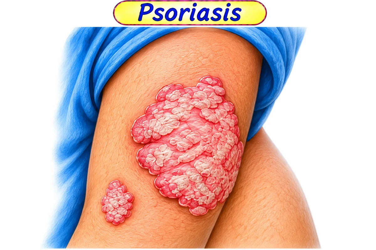
Pathophysiology: Psoriasis is a chronic, non-contagious autoimmune dermatological disease mediated by T-helper cells (Th1 and Th17). Activated T-lymphocytes release inflammatory cytokines (TNF-α, IL-17, IL-23) that trigger hyperproliferation of epidermal keratinocytes. Normal epidermal renewal cycle shortens drastically from the standard 28 days down to just 3–4 days. Immature nucleated keratinocytes accumulate rapidly, forming characteristic thick, erythematous plaques covered with silvery-white micaceous scales (Auspitz's sign: pinpoint bleeding upon scale removal).
2. Dermatitis (Eczema)
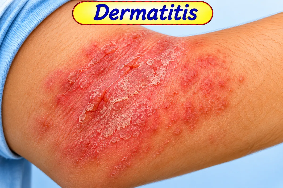
Pathophysiology: Dermatitis denotes non-specific inflammation of the epidermis and dermis characterized by an itchy, pruritic response. It comprises:
- Atopic Dermatitis: Type I IgE-mediated hypersensitivity associated with genetic epidermal barrier defects (filaggrin mutation), asthma, and allergic rhinitis.
- Contact Dermatitis: Either irritant-induced (direct chemical trauma from soaps/solvents) or allergic (Type IV cell-mediated delayed hypersensitivity to nickel, poison ivy, or topical antibiotics).
3. Leprosy (Hansen's Disease)
Pathophysiology: Leprosy is a chronic granulomatous mycobacterial infection caused by Mycobacterium leprae (and Mycobacterium lepromatosis). The acid-fast bacillus has a remarkable tropism for macrophages in the cool peripheral tissues of the skin and Schwann cells of peripheral nerves (ulnar, median, peroneal, and greater auricular nerves).
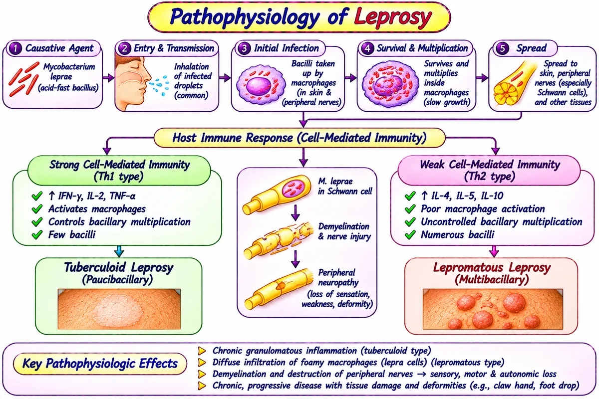
Ridley-Jopling Clinical Spectrum:
Strong cell-mediated immunity (Th1). Few hypopigmented anaesthetic skin macules with distinct margins; localized severe nerve enlargement; low bacillary load (Paucibacillary).
Depressed cell-mediated immunity (Th2). Innumerable nodular diffuse skin lesions, "leonine facies", widespread sensory loss, resorption of digits; high bacillary load (Multibacillary).
2. Basic Principles and Stages of Wound Healing
Simple Definition:
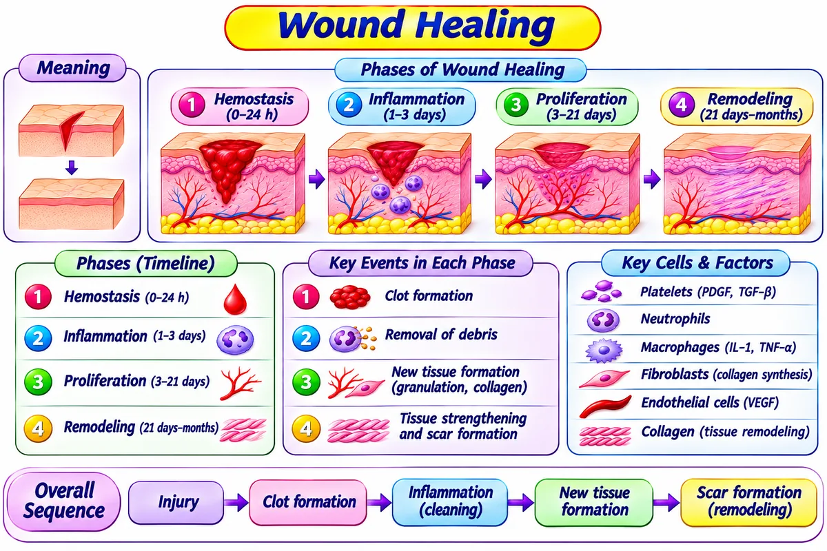
Wound Healing is defined as the biological process by which the skin and damaged tissues repair themselves after trauma, incision, or injury.
- • It is also known as: Tissue repair and regeneration.
- • It is responsible for: Restoring tissue integrity, preventing microbial infection, and stopping blood loss.
- • It is of 4 sequential stages: 1) Hemostasis, 2) Inflammation, 3) Proliferation (granulation), and 4) Remodeling (maturation).
Visual Diagram: The 4 Chronological Phases of Wound Healing
Vascular vasoconstriction; platelet plug forms; fibrin mesh seals the wound into a protective scab.
Neutrophils and macrophages phagocytose bacteria, dead debris, and release growth factors.
Fibroblasts deposit collagen; angiogenetic budding forms red vascular granulation tissue; re-epithelialization closes surface.
Type III collagen is replaced by strong Type I collagen along lines of tension; scar shrinks and regains ~80% tensile strength.
| Feature | Primary Intention (First Intention) | Secondary Intention (Second Intention) |
|---|---|---|
| Wound Nature | Clean, surgical incision with closely opposed edges sutured together. | Large, jagged wound with extensive tissue loss (severe ulcer, burns, infected wound). |
| Granulation Tissue | Minimal amount formed. | Abundant exuberant granulation tissue fills defect from bottom upwards. |
| Healing Speed & Scar | Rapid healing; leaves a fine, neat hairline scar. | Very slow healing; wound contraction by myofibroblasts leaves a large prominent scar. |
3. Skeletal System: Axial and Appendicular Divisions (206 Bones)
Simple Definition:
The Human Skeletal System is defined as the mineralized internal framework consisting of exactly 206 bones in an adult, supporting the body, protecting delicate organs, storing calcium/phosphate, and manufacturing blood cells via red marrow hematopoiesis.
A. Axial Skeleton (80 Bones)
Forms the central vertical longitudinal axis of the human body:
• Skull (22 bones): 8 Cranial bones (protect brain) + 14 Facial bones.
• Associated Bones (7 bones): 6 Auditory ossicles (Malleus, Incus, Stapes) + 1 Hyoid bone.
• Vertebral Column (26 bones): 7 Cervical, 12 Thoracic, 5 Lumbar, 1 Sacrum (5 fused), 1 Coccyx (4 fused).
• Thoracic Cage (25 bones): 1 Sternum (breastbone) + 24 Ribs (12 pairs: 7 true, 3 false, 2 floating).
B. Appendicular Skeleton (126 Bones)
Comprises the upper and lower limbs and their supporting limb girdles:
• Pectoral (Shoulder) Girdle (4 bones): 2 Clavicles (collarbones) + 2 Scapulae (shoulder blades).
• Upper Limbs (60 bones): 2 Humerus, 2 Radius, 2 Ulna, 16 Carpals (wrist), 10 Metacarpals, 28 Phalanges.
• Pelvic (Hip) Girdle (2 bones): 2 Coxal (hip) bones (Ilium, Ischium, Pubis).
• Lower Limbs (60 bones): 2 Femurs (thigh - longest bone), 2 Patellae (kneecaps), 2 Tibiae, 2 Fibulae, 14 Tarsals (ankle), 10 Metatarsals, 28 Phalanges.
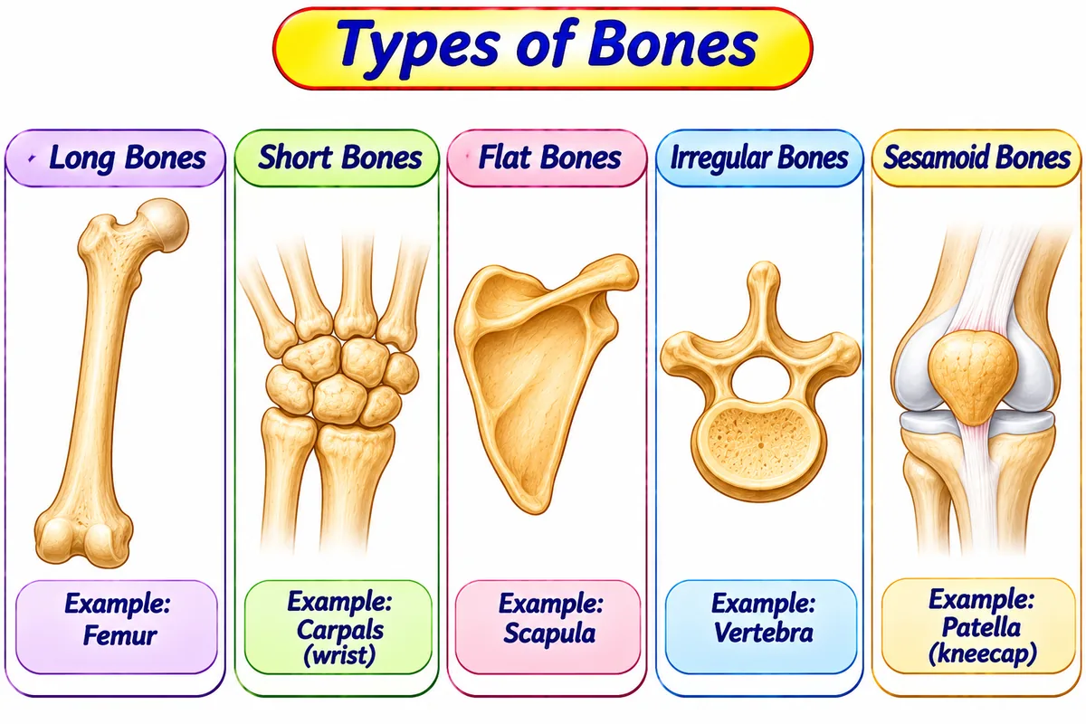
Types of Bones Classified by Shape:
4. Muscle Physiology: Sliding Filament Theory & Neuromuscular Junction
The Neuromuscular Junction (NMJ) Transmission:
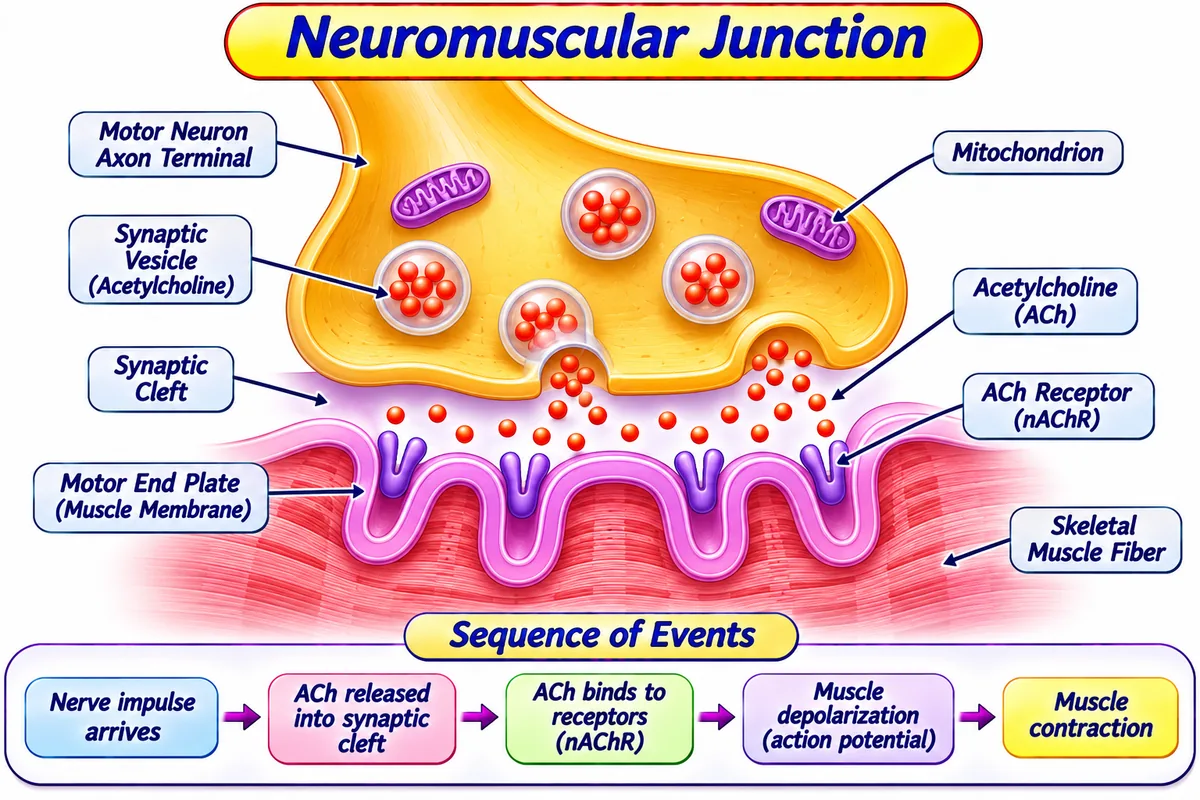
The NMJ is defined as the specialized synapse where a somatic motor nerve terminal communicates with the motor end-plate of a skeletal muscle fiber.
The Sliding Filament Theory of Muscle Contraction (Huxley):
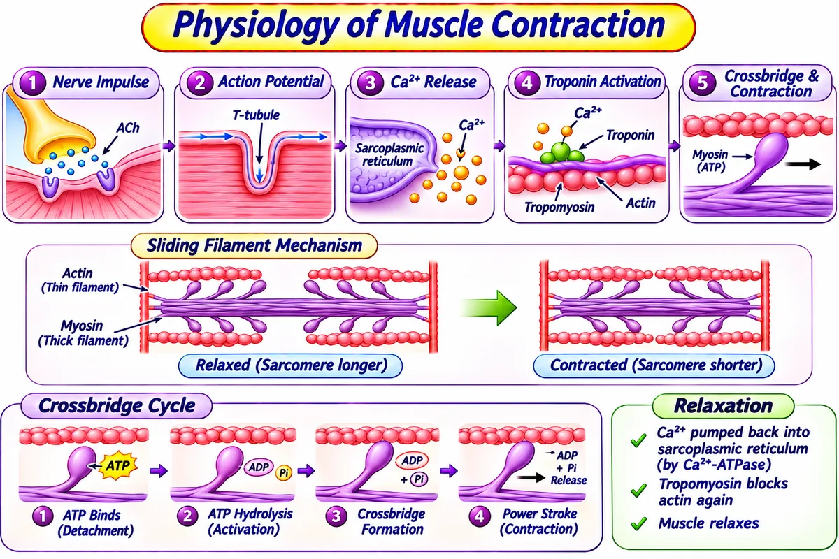
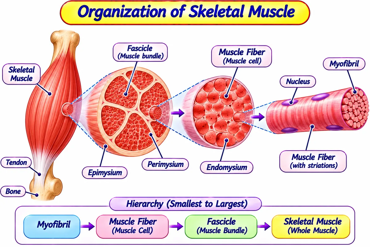
During contraction, muscle fibers do NOT shorten their filaments; rather, the thin Actin filaments slide inward past the thick Myosin filaments, shortening the sarcomere (Z-lines pull closer):
1. Calcium Binding: Intracellular Ca²⁺ binds to Troponin C, shifting Tropomyosin aside and uncovering the myosin-binding sites on Actin.
2. Cross-Bridge Formation: Energized Myosin heads bind firmly to exposed Actin.
3. The Power Stroke: Myosin heads swivel 45 degrees, pulling actin toward the M-line of sarcomere; ADP and inorganic phosphate (Pi) are released.
4. Cross-Bridge Detachment: A fresh molecule of ATP binds to myosin head, releasing it from actin (without ATP, muscles lock permanently in Rigor Mortis after death).
5. Relaxation: Active Ca²⁺-ATPase pumps return calcium back into sarcoplasmic reticulum.
5. Structural and Functional Classification of Joints
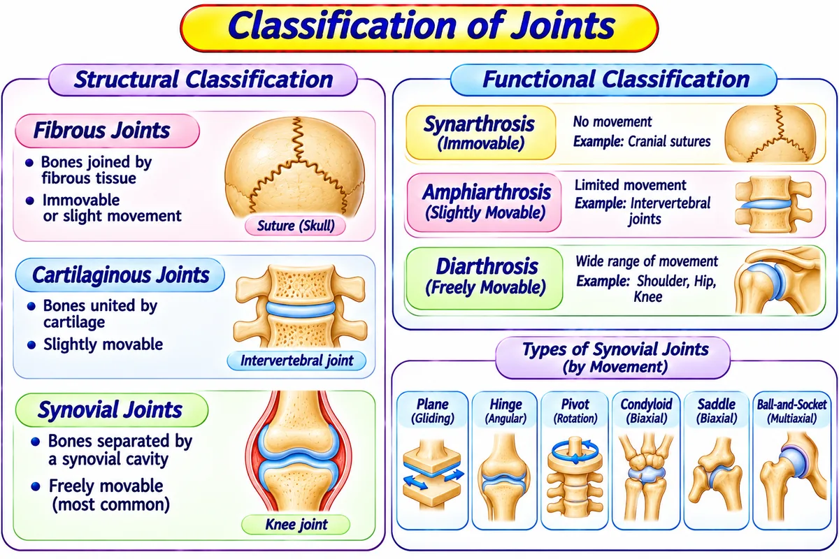
An Articulation (Joint) is defined as a point of junction between two or more bones, between bone and cartilage, or between bone and teeth.
| Structural Class | Functional Class (Mobility) | Anatomical Features | Human Body Examples |
|---|---|---|---|
| 1. Fibrous Joints | Synarthrosis (Immovable) | Bones joined directly by dense fibrous connective tissue; no joint cavity. | Sutures between cranial bones of skull; Gomphosis (teeth in jaw sockets). |
| 2. Cartilaginous Joints | Amphiarthrosis (Slightly Movable) | Bones connected tightly by hyaline cartilage or fibrocartilage pads; no cavity. | Pubic symphysis; Intervertebral discs of spinal column. |
| 3. Synovial Joints | Diarthrosis (Freely Movable) | Contains synovial cavity lined by synovial membrane secreting lubricating synovial fluid; articular hyaline cartilage over ends. | Ball-and-Socket (Shoulder, Hip); Hinge (Knee, Elbow); Pivot (Atlas-Axis). |
6. Pathophysiology of Bone and Joint Diseases: RA, Osteoporosis & Gout
A. Rheumatoid Arthritis (RA)
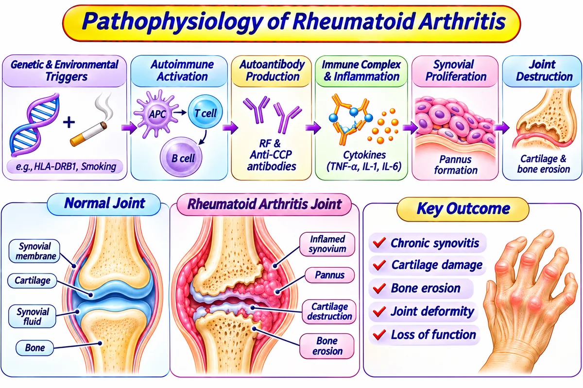
Rheumatoid Arthritis is defined as a chronic systemic autoimmune inflammatory disorder characterized by symmetric destruction of synovial joints.
• It is also known as: Autoimmune inflammatory polyarthritis.
• It is used for: Distinguishing autoimmune erosive joint damage from degenerative mechanical osteoarthritis.
• In this mechanism, we: See autoantibodies (RF and anti-CCP) trigger synovial pannus formation that degrades articular cartilage and bone.
• For example: Morning joint stiffness lasting over 1 hour affecting fingers and wrists symmetrically.
Pathophysiology: Rheumatoid arthritis is a chronic, systemic autoimmune inflammatory disease primarily targeting synovial joints. Autoantibodies—notably Rheumatoid Factor (RF) (an IgM directed against IgG Fc fragments) and Anti-Citrullinated Protein Antibodies (ACPA / Anti-CCP)—trigger immune complex deposition within the synovial membrane.
B. Osteoporosis
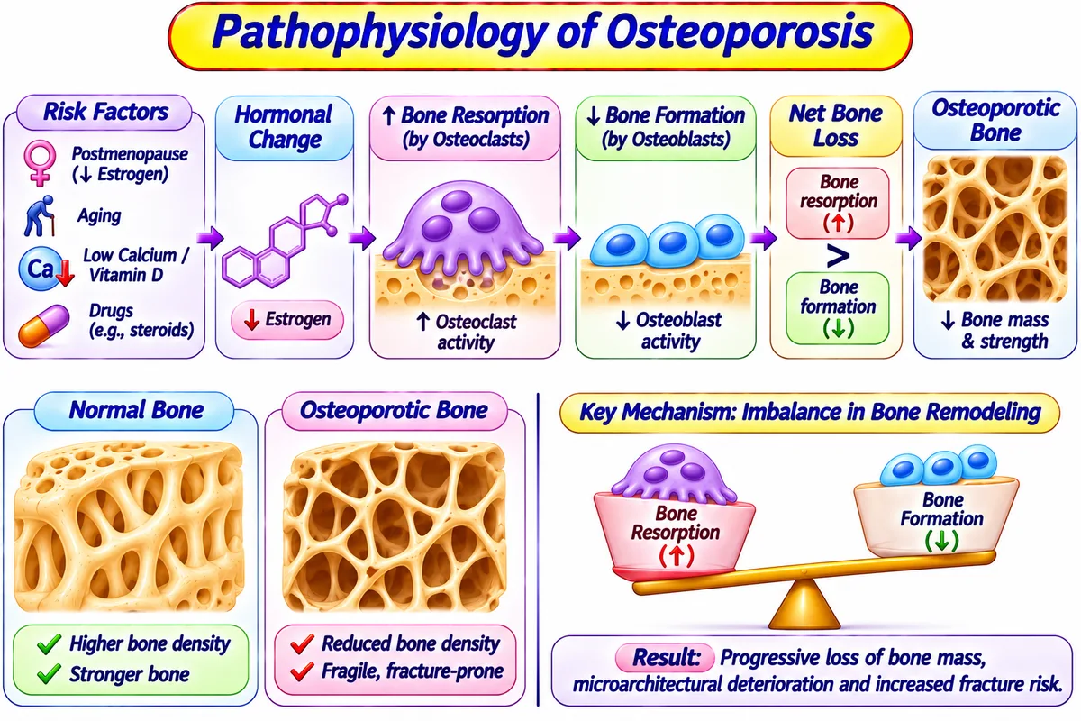
Osteoporosis is defined as a progressive metabolic skeletal disorder characterized by low bone mineral density and porous, brittle bone architecture.
• It is also known as: The silent bone thief.
• It is used for: Identifying high fracture vulnerability in postmenopausal women and the elderly.
• In this mechanism, we: See osteoclast-mediated bone breakdown exceed osteoblast-mediated bone rebuilding.
• For example: Hip fracture or vertebral compression fracture after a minor slip.
Pathophysiology: Osteoporosis is a progressive systemic metabolic skeletal disorder characterized by low bone mass and microarchitectural deterioration of bone tissue. In normal bone remodeling, bone resorption by Osteoclasts is balanced by bone deposition by Osteoblasts. In osteoporosis, resorption heavily exceeds formation, resulting in thinned cortical bone and porous trabeculae with high fracture susceptibility.
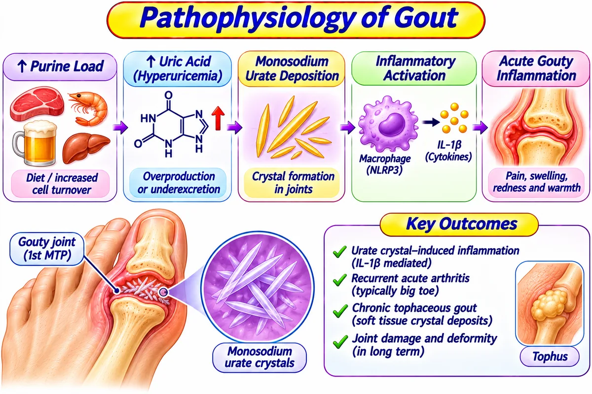
C. Gout (Gouty Arthritis)
Gout is defined as a metabolic disorder of purine catabolism characterized by hyperuricemia and precipitation of monosodium urate crystals in synovial joints.
• It is also known as: Uric acid crystal arthritis or Podagra.
• It is used for: Explaining sudden, excruciating monoarticular joint flares triggered by crystal deposits.
• In this mechanism, we: See serum uric acid exceed 6.8 mg/dL, causing needle-shaped crystals to recruit neutrophils and trigger severe inflammation.
• For example: Waking up with severe swelling, redness, and agonizing pain in the big toe.
Pathophysiology: Gout is a metabolic purine metabolism disorder characterized by persistent Hyperuricemia (serum uric acid concentration > 6.8 mg/dL). When saturation limits are exceeded, monosodium urate (MSU) crystals precipitate in avascular or low-temperature tissues such as synovial fluid, cartilage, and periarticular bursae.